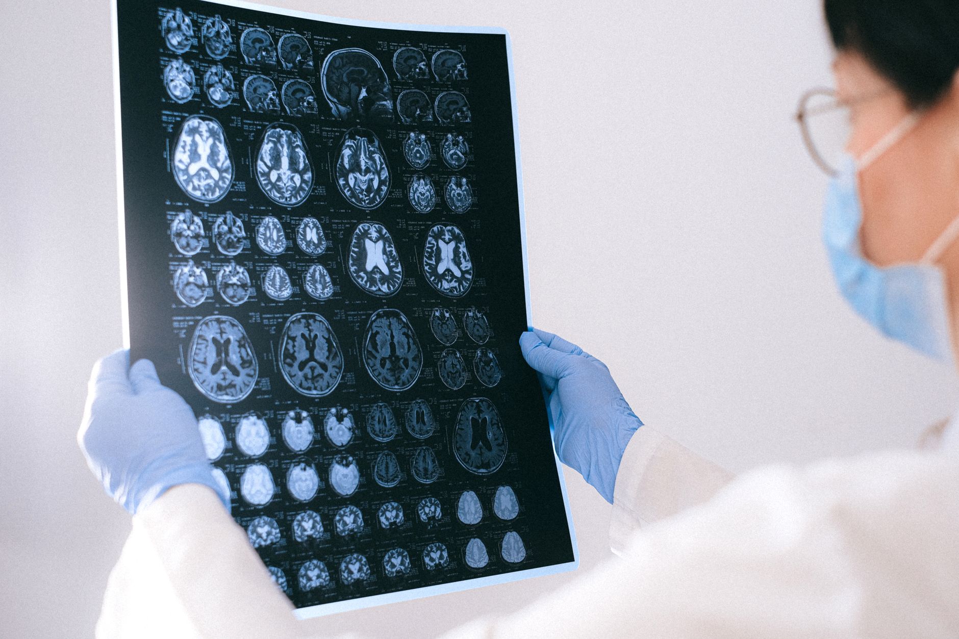
An artificial intelligence (AI) tool developed by researchers at NYU Langone Health can potentially predict a woman’s risk of developing breast cancer years before a diagnosis occurs, according to a new study. By analyzing longitudinal data from thousands of 3D mammograms, the model identifies subtle changes in breast tissue that traditional screening methods might overlook.
Key Takeaways
- Predictive Window: The AI model is designed to estimate a woman’s risk of developing breast cancer within a five-year timeframe.
- Massive Dataset: Researchers trained the model using more than 313,000 mammograms collected from over 161,000 women.
- Longitudinal Analysis: Unlike standard screenings that look at a single point in time, this tool tracks changes in breast tissue across several years of 3D mammography.
- Personalized Screening: The technology could allow doctors to tailor screening protocols, such as recommending MRIs for high-risk patients.
- Current Status: The model is currently a research tool and has not yet received FDA authorization for clinical use.
- Supplemental Imaging: Moving beyond standard mammograms to include MRI scans.
- Increased Frequency: More frequent monitoring to catch changes as they happen.
- Risk-Reduction Strategies: Early discussions regarding preventative medications or surgical options.
- If you have dense breasts: You may benefit from AI tools that help differentiate between benign tissue density and actual risk factors, potentially reducing the number of false positives you encounter.
- If you have a family history: AI can serve as a powerful supplement to your genetic testing, looking for biological markers in your tissue that genetics alone might not reveal.
- If you are concerned about radiation: One of the primary advantages of this specific AI model is that it analyzes mammograms a woman has already had. This means the risk assessment does not require additional imaging exams or extra radiation exposure.
- Multi-manufacturer validation studies to ensure technological neutrality.
- FDA regulatory filings regarding the model’s predictive accuracy.
- Clinical implementation trials that measure whether AI-guided screening actually improves long-term patient outcomes.
- www.foxnews.com
What Happened
Researchers at NYU Langone Health and its Perlmutter Cancer Center have developed an artificial intelligence model capable of estimating a woman’s risk of developing breast cancer within the next five years. The findings, published in the American Journal of Roentgenology, represent a significant shift from reactive screening to proactive risk assessment.

The development of this tool relied on an extensive dataset comprising more than 313,000 mammograms. These scans were provided by over 161,000 women, allowing the AI to learn the intricate patterns and physiological shifts that precede the development of malignant tumors.
While traditional mammography focuses on identifying existing tumors, this AI-driven approach looks backward and forward. It examines several years of a woman’s 3D mammograms to monitor how her breast tissue evolves. By identifying these specific patterns of change, the model provides a risk score that can inform future medical decisions.
Why It Matters
For decades, breast cancer screening has relied heavily on detecting visible abnormalities. However, many women receive a diagnosis despite having no known genetic predisposition or family history of the disease. This gap in traditional risk assessment highlights the need for more nuanced diagnostic tools.
According to Connie Lehman, a breast imaging specialist and professor of radiology at Harvard Medical School, the ability to identify high-risk women before cancer becomes visible is a critical advancement. Lehman, who was not involved in the study, noted that many patients express shock upon diagnosis because they lack the typical risk factors like family history or specific genetic mutations.
Furthermore, the technology addresses a major public health challenge: screening adherence. Current data suggests that between 30% and 40% of women do not participate in regular mammogram screenings. By providing more personalized and potentially less frequent or more targeted screening schedules, AI tools may help increase engagement with preventative healthcare.
The Science of AI Risk Modeling
To understand how this tool functions, it is necessary to distinguish between standard 2D mammography and the 3D mammography AI approach used in this research. Traditional 2D mammograms provide a flat image of the breast, which can sometimes result in overlapping tissue masking potential tumors. 3D mammography, or digital breast tomosynthesis, creates multiple thin slices of the breast, providing a much clearer view of the internal structures.

The NYU Langone model takes this a step further by employing longitudinal analysis. Rather than treating each mammogram as an isolated event, the AI treats a woman’s imaging history as a continuous narrative. It looks for “hidden clues”—subtle shifts in tissue density, architecture, or texture that occur over several years. These shifts may not be large enough to trigger a human radiologist’s suspicion of cancer today, but they may indicate a trajectory toward malignancy in the future.
Comparing Risk Assessment Methods
| Feature | Traditional Risk Assessment | AI-Driven Risk Assessment |
|---|---|---|
| Primary Data Points | Family history, genetic mutations (e.g., BRCA) | Longitudinal 3D mammogram tissue patterns |
| Temporal Focus | Static/Historical data | Changes in tissue over several years |
| Clinical Utility | General risk categorization | Personalized, individualized screening schedules |
| Requirement | Genetic testing or detailed family history | Analysis of existing imaging scans |
Stakeholder Perspectives and Clinical Impact
Medical experts suggest that the integration of mammogram AI technology could fundamentally change the conversation between doctors and patients. Instead of a binary “clear” or “not clear” result, patients could receive a nuanced risk profile.
Dr. Elisa Port, Mount Sinai’s Breast Surgery Director and author of ‘The Breast Advice’, has noted that AI can assist in uncovering hidden tumors and significantly reducing false positive results. This is particularly vital for women with dense breast tissue, a known complicating factor in breast cancer detection. Dense tissue can appear white on a mammogram, much like a tumor, which often leads to diagnostic uncertainty.
For high-risk individuals identified by the AI, the clinical path might include:
Conversely, for women identified as being at average risk, the tool could validate the continuation of routine, standard mammography, potentially preventing unnecessary anxiety or invasive follow-up procedures.
What It Means for You
If you are a patient currently undergoing breast cancer screenings, the emergence of this technology suggests a shift toward personalized medicine.
As these tools move from research to the clinic, patients should expect more data-driven discussions with their healthcare providers regarding exactly how often and by what method they should be screened.
Counterpoints and Open Questions
Despite the promise of the NYU Langone model, several significant hurdles remain before it can be implemented in routine clinical practice.
First, the tool is currently a research tool and is not FDA-authorized for clinical use. This means doctors cannot yet use the AI risk score to make official medical decisions or change treatment plans. The transition from a controlled research environment to a diverse, real-world clinical setting is often a rigorous and lengthy process.
Second, there are concerns regarding the generalizability of the study. The researchers noted that the current model was trained using mammograms from NYU Langone hospitals and imaging equipment from a single manufacturer. Because different mammography machines can produce slightly different image qualities and textures, the AI may need to be recalibrated to ensure it works accurately across different brands and hospital systems.
Finally, it is crucial to remember that an AI risk score is not a cancer diagnosis. As Connie Lehman cautioned, a higher risk score does not mean a woman has cancer, nor does a lower score guarantee that she is safe. The tool is a predictive indicator, not a definitive proof of disease.

What Happens Next
The research team at NYU Langone has already outlined the next steps for validating this technology. To ensure the model is robust and applicable to the general population, they plan to conduct further testing using data from other medical centers and across different 3D mammography systems.
Success in these multi-center trials will be the primary catalyst for seeking FDA authorization. Once authorized, the tool could be integrated directly into radiology workflows, providing real-time risk assessments alongside standard diagnostic reports.
Watch for developments in:
Frequently Asked Questions
How does AI predict breast cancer risk differently than a doctor?
A traditional radiologist looks at a single mammogram to see if a tumor is present now. The AI model used by NYU Langone looks at a series of mammograms over several years. It uses machine learning to identify subtle, longitudinal changes in the pattern and density of breast tissue that are too minute for the human eye to track consistently over time.
Will I need more mammograms to get this AI risk score?
No. One of the key benefits of this technology is that it can analyze the mammograms you have already undergone. It uses the existing data in your medical history to generate a risk assessment, meaning it does not inherently require additional radiation or extra imaging sessions.
Is an AI risk score the same as a cancer diagnosis?
No. It is critical to understand that a high risk score is a predictive indicator, not a diagnosis. It suggests a higher probability of developing cancer in the next five years, which may prompt a doctor to suggest more frequent or more detailed screenings, such as an MRI. It does not mean cancer is currently present.
Why is the study’s use of a single manufacturer a concern?
Medical imaging equipment can vary between manufacturers. A model trained exclusively on images from one brand of machine might become “accustomed” to the specific digital signatures or textures produced by that brand. To be useful in a general hospital setting, the AI must prove it can accurately interpret images from all types of 3D mammography systems.
Closing
The development of the NYU Langone AI model marks a significant step toward a future where breast cancer screening is as much about prediction as it is about detection
References
Featured image: Photo by Anna Shvets on Pexels