
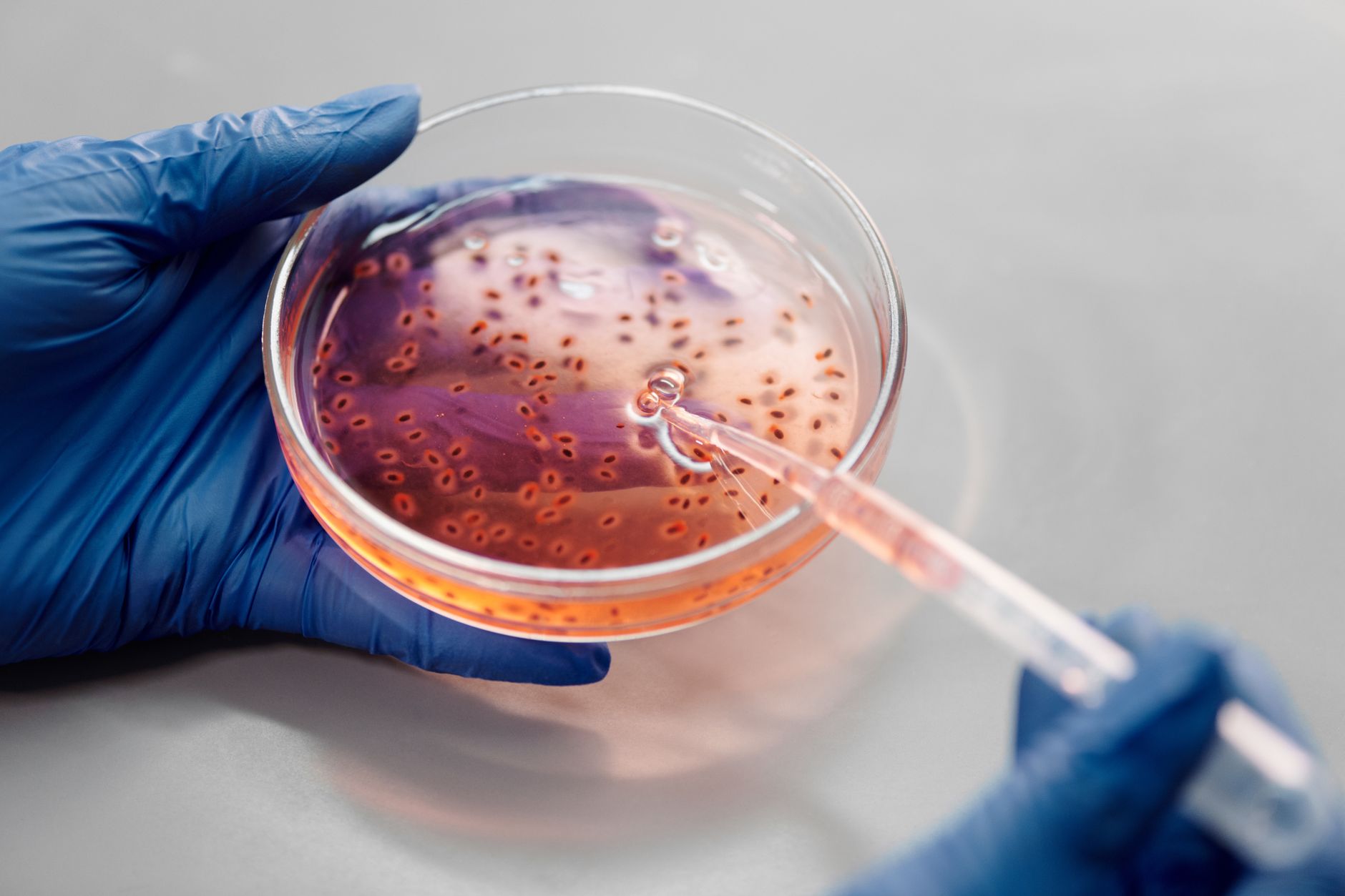
The integration of human brain organoids into the brains of mice marks a significant leap in the quest to model complex human neurological processes within a living organism. Researchers at Stanford University have successfully replaced a substantial portion of the mouse cortex with human tissue, providing a potential new framework for studying how human cells interact within a functional nervous system. This breakthrough, detailed in a study published in the journal Nature in 2026, attempts to bridge the gap between isolated laboratory tissue and the complex, interconnected reality of a living brain.
Key Takeaways
- Successful Integration: Approximately 85% of the mice implanted with human organoids successfully incorporated the graft, with human cells making up 92% of the resulting cortex.
- Functional Connectivity: Human neurons formed long-distance connections, with cellular processes detected as far away as the spinal cord.
- Behavioral Improvements: Mice with humanized cortices showed intermediate performance in motor coordination and weight compared to normal and cortex-free mice.
- Structural Limitations: Despite cellular integration, the humanized tissue lacked the distinct, organized layers characteristic of a healthy, natural cortex.
- Model Potential: While not yet a perfect model for complex diseases like ALS, the system shows promise in observing how human cells respond to physiological stressors like hypoxia.
- For Medical Researchers: This provides a new, albeit imperfect, model to study human-specific responses to stimuli, such as hypoxia (oxygen deprivation), which the study noted human cells responded to in ways consistent with normal human biology.
- For Pharmaceutical Developers: If these models can be refined to include organized layers, they could become essential for testing the efficacy and toxicity of drugs targeting human-specific neural pathways before moving to human clinical trials.
- For Patients with Neurological Diseases: While this is basic science, the long-term goal is to use such models to understand diseases like ALS, Alzheimer’s, or Parkinson’s in a way that more accurately reflects human biology than current animal models.
- For Biotech Investors: The success of this “space-clearing” technique suggests a new frontier in neuro-engineering, potentially opening up new avenues for research into brain-machine interfaces and regenerative therapies.
- Detailed Anatomical Mapping: Using advanced imaging to understand the specific connections being made and why the layered structure is failing to form.
- Functional Quantification: Determining exactly which neurological tasks are being aided by the human cells and which remain unaddressed.
- Refining the Environment: Investigating ways to provide the human organoids with better structural cues—perhaps through more advanced scaffolds or chemical signaling—to encourage the formation of organized cortical layers.
- Disease Modeling: Once the structural issues are addressed, the team can begin introducing specific disease markers into the human organoids to see how they behave within the living mouse model.
- arstechnica.com
What Happened
In a study reported by John Timmer for Ars Technica and published in Nature on September 16, 2026, a research group at Stanford University described a method to bypass the inherent limitations of traditional brain organoids. For years, scientists have used organoids—small, three-dimensional patches of tissue grown from stem cells—to study human diseases. However, these organoids typically exist in isolation, lacking the circulatory systems, immune cells, and long-range neural connections found in a complete organism.
To address this, the Stanford team took the radical step of genetically removing the cortex from mice and replacing it with human brain organoid cells. The researchers identified a gene active in nearly all cortical cells and utilized it to trigger the deletion of a critical gene responsible for chromosome separation during cell division. This process effectively prevented the development of a natural mouse cortex, reducing the brain’s volume by half.
To ensure the survival of the mice, which would otherwise succumb to the loss of their cortex, the team implemented intensive care protocols. According to the study, researchers had to eliminate most of the pups in a litter to ensure the cortex-free mice received adequate nursing. The surviving mice were allowed to nurse longer and were subsequently provided with high-calorie food to maintain their health. Additionally, the mice were immunocompromised to prevent the host’s immune system from rejecting the human cellular grafts.
Why It Matters
This research matters because it moves the study of human neurology from a static petri dish into a dynamic, living environment. Traditionally, researchers have faced a binary choice: study human cells in a dish (which lacks context) or implant human stem cells into a mouse brain (where the host’s healthy neurons often mask the unique behaviors of the human cells).
By clearing out the host’s cortical space, the Stanford team created a “vacancy” that allowed human cells to take center stage. This provides a unique window into how human neurons communicate with non-human structures, such as the spinal cord, and how they might respond to the systemic environment of a living body. For the billions of people affected by neurological disorders, this model could eventually become a primary tool for testing how human-specific brain structures react to drugs or disease progression.
The Biological Challenge of Human-Mouse Integration
Integrating human tissue into a different species is fraught with biological hurdles. One of the primary issues is the vastly different developmental timelines between humans and mice. Human neurons mature significantly more slowly than those of mice. In a natural mouse pregnancy, which lasts only about 21 days, human cells might not have enough time to form the connections required for the animal to survive or function.
Furthermore, the lack of organized structure in the resulting humanized brain highlights a fundamental problem in regenerative medicine: presence does not equal organization. While the human cells successfully occupied the space and formed various neuron types, they did not replicate the highly structured, layered architecture of a natural cortex. This suggests that while the “building blocks” are present, the “blueprints” provided by the host environment may be insufficient to guide human cells into complex, organized patterns.
Comparative Data of Cortical Replacement Outcomes
To understand the impact of the humanized cortex, the researchers compared three distinct groups of mice: normal mice, mice with no cortex, and mice with the humanized cortex graft.
| Feature | Normal Mice | Cortex-Free Mice | Humanized Mice (Human Organoids) |
|---|---|---|---|
| Cortex Structure | Organized, distinct layers | None | Disorganized; no distinct layers |
| Cellular Composition | 100% Mouse neurons | 0% Cortex | ~92% Human cells |
| Body Weight | Normal/Standard | Significantly lighter | Intermediate |
| Maze Navigation | High accuracy | Random chance | Better than chance; below normal |
| Motor Coordination | Normal | Poor | Intermediate |
| Associative Memory | High capability | No capability | No significant improvement |
Deep-Dive: Behavioral and Physiological Analysis
To assess how the humanized cortex affected the mice, the researchers utilized a machine-learning classifier to analyze video monitoring of the animals’ behaviors. This allowed them to categorize movement patterns into distinct clusters. The results confirmed that the humanized mice occupied a unique neurological space, distinct from both healthy mice and those without a cortex.
While the humanized mice performed better than those with no cortex in certain areas, such as fine motor coordination and weight maintenance, they did not reach the level of performance seen in normal mice. Specifically, in simple maze tests, the humanized mice performed better than random chance, yet they failed to show improvement in associative memory tests. This indicates that while the human cells provide some level of functional support, they have not yet replicated the higher-order cognitive processing capabilities of a natural cortex.
What It Means for You
For different stakeholders, the implications of this research vary significantly:
Counterpoints and Open Questions
Despite the excitement, several critical questions and risks remain. The most prominent issue is the lack of large-scale organization. Without the distinct layers of the cortex, it is difficult to determine if the human cells are truly performing “human-like” functions or if they are merely acting as a biological filler.
Critics and cautious observers might argue that a disorganized mass of human neurons is not a sufficient proxy for the human brain. There is a risk that the connections formed—while present—are too chaotic to provide meaningful data on complex cognitive functions. Furthermore, the study notes that it is currently impossible to associate the mild improvements in the mice with any specific function provided by the human cells, as the anatomical structure remains uncharacterized.
Another concern involves the ethical and biological implications of creating “humanized” animals. While the mice in this study were primarily used to test the feasibility of the graft, the increasing ability to integrate human neural tissue into animals raises significant questions about the boundaries of species and the potential for unintended cognitive changes in laboratory subjects.
What Happens Next
Moving forward, the research team must move beyond proving that the cells can survive and begin quantifying what they are doing. The next phase of research will likely focus on:
Frequently Asked Questions
What are brain organoids?
Brain organoids are tiny, three-dimensional structures grown in a laboratory from stem cells. They are designed to mimic the complexity of human brain tissue by allowing cells to self-organize into various types of neurons and rudimentary structures. Unlike traditional 2D cell cultures, organoids provide a more realistic environment for studying how cells interact in three dimensions.
How did researchers replace the mouse cortex?
The researchers used genetic engineering to target the cells that would normally form the cortex. By driving the deletion of a key gene necessary for proper cell division, they prevented the mouse’s own cortical cells from developing. This created a void in the brain that could then be filled by implanting human brain organoids.
Can these mice think like humans?
No. While the mice showed some improvements in behavior and motor skills compared to mice without a cortex, they did not exhibit human-like intelligence or complex cognition. The human cells in the mice were disorganized and lacked the structural complexity required for high-level brain function. The study showed they were “intermediate” between normal mice and cortex-free mice, not human-like.
Why was the study published in Nature?
Nature is one of the world’s most prestigious scientific journals. The publication of this study suggests that the methodology—specifically the ability to successfully integrate human tissue into a living host and maintain the host’s survival—is considered a significant and rigorous scientific achievement.
The Stanford study represents a vital, if early, step in the evolution of neurobiological modeling
References
Featured image: Photo by Edward Jenner on Pexels


One thought on “Human Brain Organoids: Why the Stanford Mouse Study Matters”