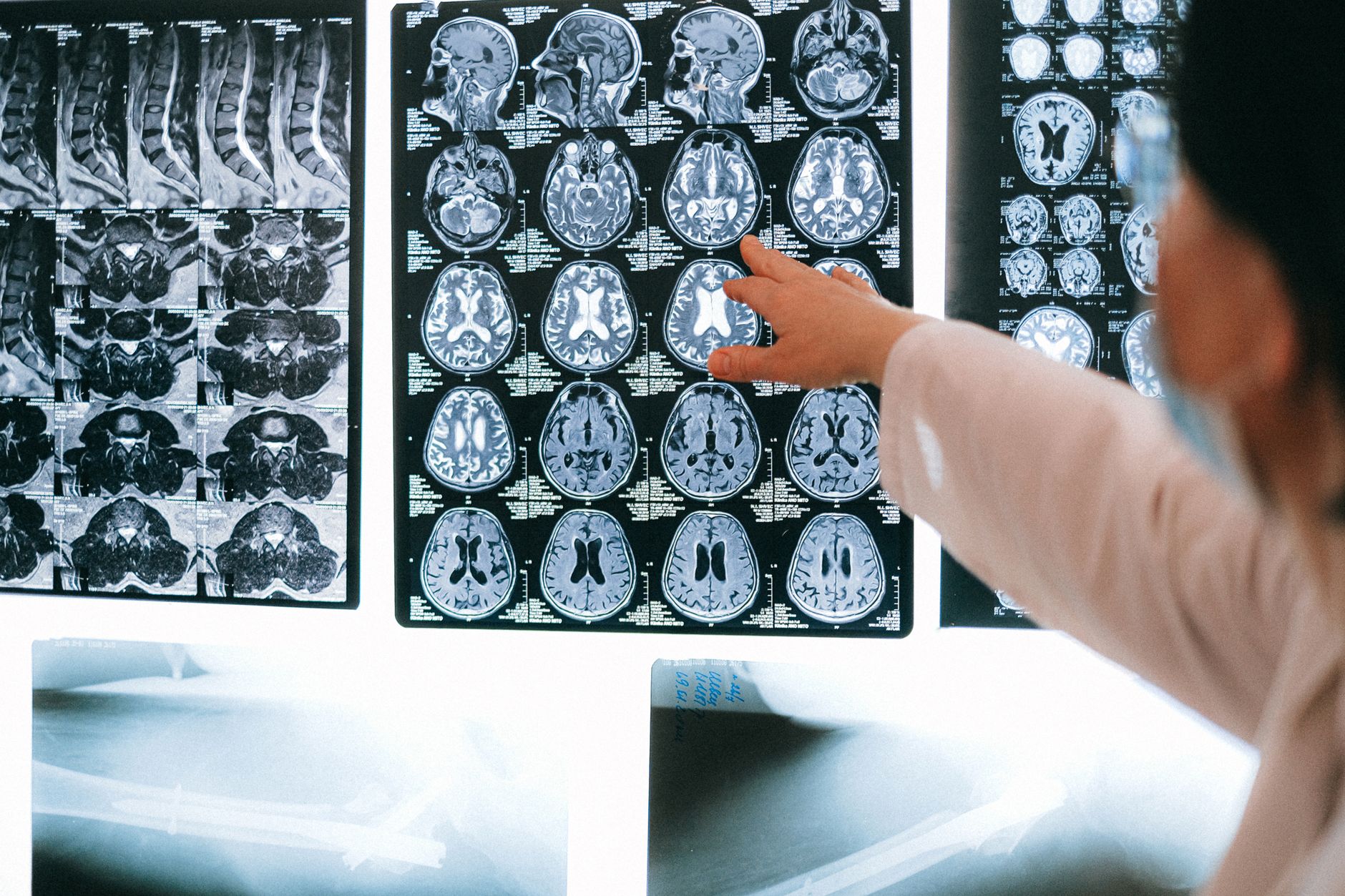
<a href="https://news.quantosei.com/2026/09/16/brain-eating-amoeba-infection-durham-water-feature-linked-to/” title=”<a href="https://news.quantosei.com/2026/09/16/brain-eating-amoeba-infection-durham-water-feature-linked-to/" title="Brain-Eating Amoeba Infection: durham water feature Linked to Death”>brain-eating amoeba infection: Durham Water Feature Linked to Death”>human brain organoids have reached a significant milestone in neurological research as scientists at Stanford University successfully integrated human tissue into the brain structure of mice to study how these cells function in a living environment.
Key Takeaways
- Successful Integration: Researchers at Stanford University successfully implanted human cortex organoids into mice that were genetically engineered to lack a natural cortex.
- High Incorporation Rate: Approximately 85% of the implanted animals successfully incorporated the human graft, which eventually accounted for 92% of the cells in the replacement cortex.
- Functional Connectivity: The human cells formed long-distance neural connections, with processes detected as far away as the spinal cord.
- Structural Limitations: While the cells integrated, they failed to form the distinct, organized layers characteristic of a healthy human or mouse cortex.
- Intermediate Cognitive Recovery: Mice with humanized tissue showed intermediate physical and cognitive performance—better than mice without a cortex, but significantly lower than normal mice.
- Modeling Potential: The study provides a foundation for future disease modeling, though it is not yet a complete solution for studying complex diseases like ALS.
- Structural Refinement: Finding ways to provide the necessary biochemical or electrical signals to encourage human organoids to form the distinct layers found in a natural cortex.
- Functional Characterization: Conducting more granular studies to determine exactly which functions are being improved by the human cells. This will involve using advanced machine-learning classifiers to analyze even more subtle behavioral patterns and neural firing sequences.
- arstechnica.com
What Happened
On September 16, 2026, a research group at Stanford University published findings describing a method to study human brain organoids within a more natural, biological context. The study, led by researchers including those from the S. Pasca lab, sought to overcome the inherent limitations of studying brain tissue in isolation.
Traditional organoids—small, three-dimensional patches of tissue grown from stem cells—often fail to mimic the complexity of a real brain because they lack connections to other specialized structures, circulatory systems, and immune cells. To address this, the Stanford team utilized a radical approach: they genetically deleted a large portion of the mouse brain—specifically the cortex—and replaced it with human brain organoid cells.
To achieve this, the researchers identified a gene active in nearly all cortical cells and used it to drive the deletion of a key gene required for chromosome separation during cell division. This process effectively prevented the mouse’s own cortex from developing. To ensure the survival of these mice, who would otherwise perish without a cortex, the team had to implement intensive care protocols, including extended nursing periods and high-calorie diets.
Why It Matters
For decades, the field of neuroscience has struggled with the “isolation problem.” While studying human cells in a petri dish is useful, those cells do not experience the long-range signaling or the systemic interactions that define human brain function.
By creating a “humanized” mouse model, scientists can observe how human neurons interact with a living nervous system. This is critical for understanding how diseases move through the brain and how they affect the body. For example, the researchers noted that the human cells responded to hypoxia (low oxygen levels) in a way that was consistent with normal human cells, rather than mouse cells. This suggests that the model can accurately reflect human-specific physiological responses, which is a vital step toward developing more effective treatments for neurological disorders.
The Challenge of Organoid Development
To understand the significance of the Stanford study, one must understand the limitations of the current state of the art. Human brain organoids are a massive leap forward from dissociated cells in a culture dish because they adopt a three-dimensional structure. However, they are essentially biological islands.
In a healthy human body, the brain is not a standalone unit. It is connected to the liver via chemical signals, to the immune system via circulating cells, and to the rest of the body via long-range axonal connections. Without these inputs, organoids cannot fully replicate the complex decision-making or sensory processing that occurs in a mature brain.
Previous attempts to bridge this gap involved implanting human neural stem cells into the brains of other species. However, because the host’s own neurons were still functioning normally, it was difficult for scientists to discern whether the observed behaviors were driven by the human cells or the host cells. The Stanford approach attempts to solve this by removing the host’s cortical influence entirely, allowing the human cells to take center stage.
Analyzing the Results: Humanized vs. Natural Mice
The researchers observed that while the human cells successfully occupied the space of the missing cortex, the resulting biological structure was significantly different from a natural brain. The human cells formed all the major types of neurons found in a cortex and even engaged in synchronized activity spikes, suggesting a level of coordination. However, the hallmark of a cortex—its distinct, organized layers—was absent.
To quantify the impact of this replacement, the team conducted a series of behavioral and physical assessments. The results showed a clear spectrum of function between the three groups of mice studied.
| Metric | Normal Mice | Cortex-Free Mice | Humanized Mice (Human Organoids) |
|---|---|---|---|
| Body Weight | Standard/Healthy | Significantly Lighter | Intermediate |
| Maze Memory | High Performance | Random Chance | Better than Chance |
| Associative Memory | High Performance | No Performance | No Performance |
| Motor Coordination | High Performance | Low Performance | Intermediate |
| Cortical Structure | Fully Layered | None | Disorganized/No Layers |

Deep-Dive: The Structural Deficit
One of the most critical findings in the Stanford brain research is the lack of large-scale organization. In a typical mammalian cortex, neurons are arranged in highly specific layers. These layers are essential for the way information is processed—for example, sensory input might enter through one layer, while motor output is processed in another.
In the humanized mice, the researchers found some evidence of local organization, meaning specific cell types tended to cluster near each other. However, the macro-structure—the “map” of the cortex—never materialized. This suggests that while human cells can integrate and communicate, they require more than just a physical space to form a functional architecture; they likely require specific developmental signals that were missing in the mouse environment.
This lack of structure explains why the mice performed better than those with no cortex but failed to reach the cognitive levels of normal mice. The “intermediate” results in weight, motor coordination, and maze navigation suggest that the human cells provided some functional benefit, but the disorganized nature of their connections prevented higher-order cognitive tasks, such as associative memory, from being mastered.
What It Means for You
While this research is currently in the laboratory phase, its implications for various stakeholders are profound:
For Medical Researchers
If you are working in neurodegenerative disease research, this study represents a potential, albeit imperfect, new tool. The ability to observe how human neurons respond to environmental stressors like hypoxia in a living organism provides a much higher level of biological relevance than current in-vitro models. As the technology for organizing these organoids improves, this could become a primary platform for drug testing.
For Patients and Families
If you or a loved one are living with conditions such as ALS, Alzheimer’s, or Parkinson’s, this research offers a glimmer of hope for the future of precision medicine. The goal of this line of inquiry is to create models that can accurately predict how a specific human patient’s brain might respond to a new medication, potentially reducing the reliance on trial-and-error treatments and accelerating the path to a cure.
For the Biotech Industry
Investors and developers in the biotechnology sector should watch the evolution of “organoid-on-a-chip” and “humanized animal” technologies. The ability to successfully graft human tissue into animal models is a high-value capability that could redefine the pharmaceutical pipeline.
Counterpoints and Open Questions
Despite the excitement, the scientific community remains cautious. A primary concern is whether the disorganized connections formed in these humanized brains will ever be sufficient to model complex human neural processing. If the cells cannot form the necessary layers, can they ever truly replicate the functions of a human brain?
Furthermore, there are significant unanswered questions regarding the long-term stability of these grafts. The researchers have not yet performed a detailed anatomical study to quantify how these structures change over the animal’s lifespan. There is also the question of individual variation: how much does the success of the graft depend on the specific batch of organoids used, and how consistent would such a model be across a large-scale study?
Finally, the use of immunocompromised mice to prevent rejection is a necessary scientific control, but it also creates a biological environment that is not entirely “natural,” which may influence the results in ways that are not yet fully understood.
What Happens Next
Moving forward, the Stanford team and other researchers in the field will likely focus on two main objectives:
Watch for upcoming publications in journals like Nature that detail the long-term survival and structural evolution of these humanized cortical grafts. The ability to bridge the gap between “cells in a dish” and “cells in a body” will be the deciding factor in whether this technology becomes a cornerstone of modern neuroscience.
Frequently Asked Questions
What are brain organoids?
Brain organoids are tiny, three-dimensional structures grown in a laboratory from human stem cells. They are designed to mimic the complexity of human brain tissue by allowing cells to self-organize into structures that resemble parts of the brain. While they are much more advanced than simple cell cultures, they currently lack the full connectivity and systemic support (like blood vessels) found in a real human brain.
How did researchers remove the mouse cortex?
According to the study, researchers used a genetic technique to target the mouse’s developing brain. They identified a specific gene that is active in almost all cortical cells and used it to trigger the deletion of a key gene responsible for separating chromosomes during cell division. This effectively prevented the mouse’s own cortical cells from maturing and forming a brain structure, creating a “vacancy” for the human organoids.
Can these humanized mice think like humans?
No. While the human cells integrated and provided some functional improvements, the mice did not exhibit human-like intelligence or complex thought. The human cells in the mice were disorganized and lacked the layered structure required for advanced cognitive processes. The mice showed improvements in basic tasks like motor coordination and simple maze navigation, but they could not perform complex associative memory tasks.
Is this a model for curing diseases like ALS?
Not yet. While the research is a significant step toward creating better disease models, the current version is not yet sophisticated enough to study complex, multi-system diseases like Amyotrophic Lateral Sclerosis (ALS). To be a useful model for ALS, the system would need to better replicate the complex interactions between neurons and the surrounding environment that drive the disease. However, it provides a much-needed foundation for reaching that goal.
Closing
The successful integration of human brain organoids into a living mouse model marks a pivotal shift in how we approach neurological research
References
Featured image: Photo by Anna Shvets on Pexels