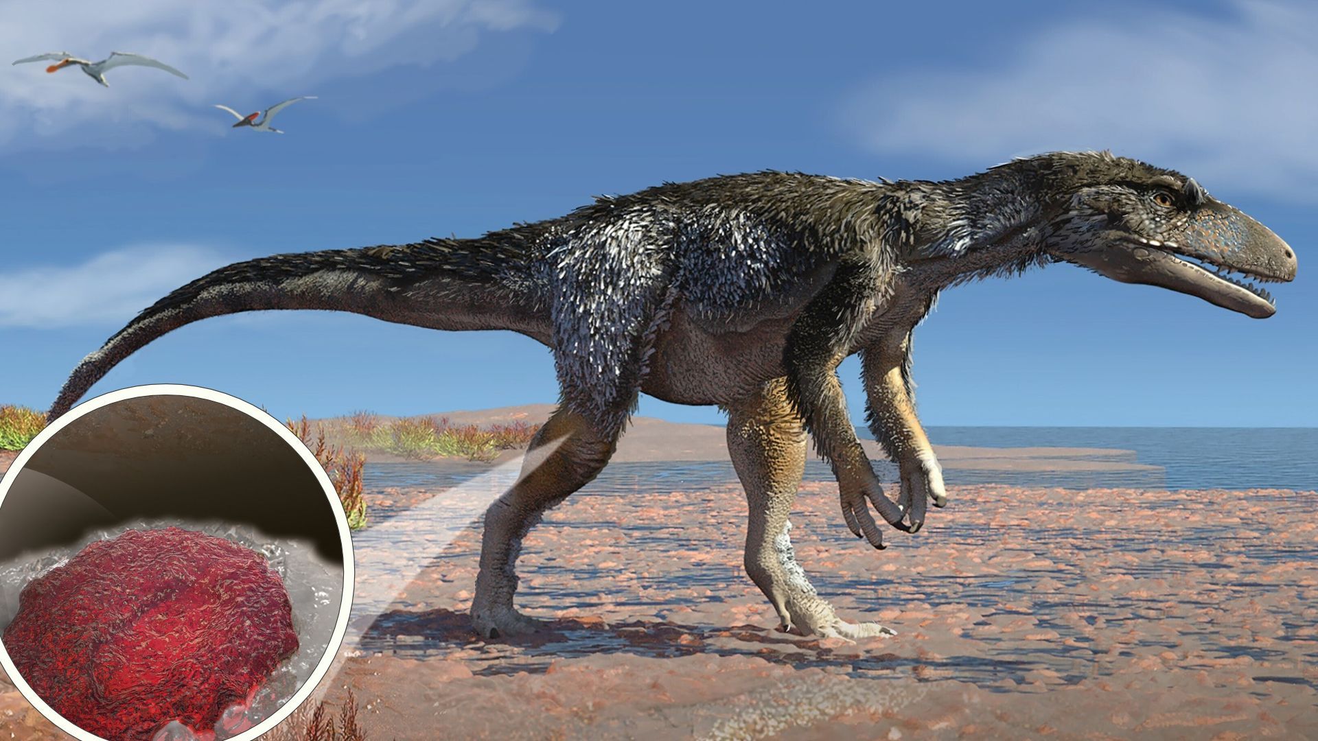
Dinosaur blood cells have been identified in a 115-million-year-old fossil, a discovery that could fundamentally change the way paleontologists study the biology of extinct species. A research team led by scientists at Curtin University in Australia has reported finding what appear to be fossilized red blood cells, alongside blood-derived molecules, preserved within the leg bone of a small theropod dinosaur.
Key Takeaways
- Major Discovery: Researchers identified microscopic, cell-like structures and heme—an oxygen-carrying molecule—inside an Aratasaurus museunacionali fossil.
- Record-Breaking Age: At 115 million years old, this represents the oldest known example of possible dinosaur blood cells, surpassing previous finds that dated to roughly 80 million years ago.
- Preservation Mechanism: Scientists believe the delicate structures were stabilized by hausmannite, a manganese oxide mineral, within a low-oxygen environment.
- Scientific Debate: While the chemical evidence is strong, experts like Mary Schweitzer question if the physical shape of the structures truly matches known non-mammalian red blood cells.
- Future Impact: The find suggests that thousands of existing museum specimens may contain untapped molecular and biological data.
- In Situ Investigation: They investigated the chemistry within the bone itself to ensure the signals were physically linked to the microstructures.
- Resonance Raman Spectroscopy: This was used to confirm the presence of specific chemical bonds.
- Elemental Mapping: This revealed that the cell-like structures were correlated with iron, carbon, and nitrogen—the primary constituents of heme. In contrast, the rest of the bone was composed of phosphorus, calcium, fluorine, and carbon.
- Microbial Exclusion: The team searched for traces of fungi, spores, or biofilms that might have colonized the bone after death, but found none, helping to rule out microbial mimicry.
- For Researchers and Students: Expect a paradigm shift in how biological data is extracted from the fossil record. The focus of study is expanding from the macro (bones) to the micro (molecules).
- For Museum Curators and Institutions: The discovery suggests that thousands of fossils currently sitting in museum drawers—collected decades ago for simple anatomical study—may actually be reservoirs of high-value molecular, isotopic, and microscopic data. This could lead to a massive re-evaluation of existing collections.
- For the Scientific Community at Large: This find challenges the “conventional wisdom” regarding the limits of organic preservation, potentially opening up new windows into the history of life on Earth.
- Are they truly cells? Could the structures be a unique type of mineral growth that merely mimics the shape of a cell?
- Is the heme truly endogenous? While the team used in situ analysis, the possibility of ancient contamination remains a central concern in molecular paleontology.
- Can this be replicated? As Grice noted, the immediate priority is replication. Scientists must determine if these signals can be found in other fossils from different geological settings and different dinosaur families.
- www.livescience.com
- www.theguardian.com
- www.sciencealert.com
What Happened
On November 2, 2026, a study published in the journal Proceedings of the National Academy of Sciences (PNAS) detailed the discovery of biological traces within the limb bones of an Aratasaurus museunacionali. The specimen, a small, meat-eating theropod dinosaur approximately 10 feet (3 meters) in length, was originally recovered from a mine in north-eastern Brazil in 2008.
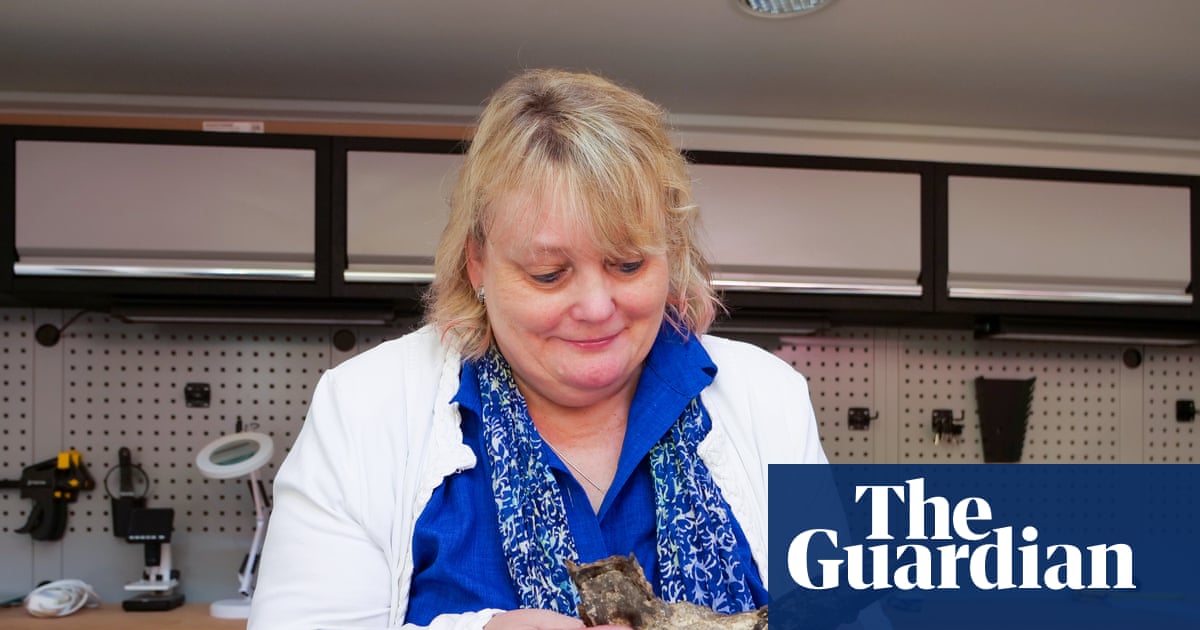
The research, led by Kliti Grice, an organic geochemist at Curtin University, focused on the fossilized vascular canals—the channels that once housed blood vessels—inside the dinosaur’s right leg bone. Upon microscopic inspection, the team discovered a collection of ellipsoidal microstructures that matched the expected size and location of red blood cells.
To move beyond mere visual observation, the team employed a rigorous multi-disciplinary approach. They utilized high-performance liquid chromatography–mass spectrometry to analyze the bone extract, which revealed a dominance of heme B. This finding was significant because the chemical signature of the heme was distinct from the surrounding rock, which appeared to have been produced by photosynthetic microbes rather than the dinosaur itself.
Why It Matters
This discovery represents a potential shift in the field of paleontology, moving the discipline from a focus on skeletal morphology—the study of bone shapes—to a more complex molecular science. If these findings are validated, scientists will no longer be limited to studying what a dinosaur looked like; they will be able to investigate how it functioned.
By accessing ancient molecular traces, researchers could potentially reconstruct the metabolism, physiology, and biochemistry of extinct animals. This would provide unprecedented clarity on the activity levels, respiratory efficiency, and even the specific blood chemistry of creatures that lived millions of years ago. Furthermore, the study suggests that the “archive” of life is much deeper than previously thought, implying that the chemical signatures of life can persist far longer than the conventional scientific consensus once suggested.
The Science of Ancient Preservation
One of the most significant hurdles in paleontology is the rapid degradation of organic matter. Traditionally, it has been believed that endogenous molecules—those produced within a living organism—disappear within a million years or less. The presence of these structures after 115 million years challenges that timeline.
The Role of Hausmannite and Mineralization
According to Kliti Grice, the preservation was likely not a matter of the original biological cells surviving unchanged, but rather a process of mineral replication. The team proposes that the dinosaur died and was quickly buried in a shallow-water environment characterized by a low-oxygen (anoxic) setting and the presence of microbial mats.
In this specific geochemical environment, a manganese oxide mineral known as hausmannite ($Mn3O4$) likely played a crucial role. The researchers suggest that this mineralization occurred early in the fossilization process, effectively “stabilizing” or replicating the delicate cellular structures before they could decompose. This created a mineralized record of the original biological structures.
Advanced Analytical Techniques
The team did not rely solely on visual morphology. To ensure the findings were not the result of post-mortem contamination, they conducted several layers of testing:
| Feature | Research Team’s Claim (Grice et al.) | Scientific Critique (Schweitzer) |
|---|---|---|
| Structure Identity | Fossilized red blood cells | Unconfirmed microstructures |
| Morphology | Ellipsoidal/Cell-like in vascular canals | Concave; lacks “nuclear bulge” |
| Size | Consistent with cell-like structures | Smaller than most non-mammalian RBCs |
| Chemical Basis | Presence of heme B and collagen amino acids | Acknowledges endogenous molecules can persist |
Stakeholders and Scientific Controversy
The study has ignited a significant debate within the paleontological community. While the technological rigor of the Curtin University team is widely respected, the biological interpretation of the findings is contested.
The Morphological Critique
Mary Schweitzer, a prominent molecular paleontologist at North Carolina State University, has expressed skepticism regarding the claim that these structures are indeed red blood cells. Her critique centers on the biological differences between mammalian and non-mammalian (avian and reptilian) blood cells.
Unlike mammalian red blood cells, which lack a nucleus, the red blood cells of birds and reptiles retain their nuclei. This creates a characteristic “nuclear bulge,” giving the cells a dome-like appearance. Schweitzer notes that the structures observed in the Aratasaurus fossil appear concave and lack this bulge. Additionally, she points out that the structures identified by the team are smaller than most known non-mammalian red blood cells.
“I am in agreement that endogenous molecules can persist over geological time in some form, and that there is much these can teach us,” Schweitzer said, acknowledging the importance of the team’s molecular approach. However, she concluded that “morphological similarity is not met.”
The Research Team’s Defense
Grice and her colleagues maintain that the convergence of independent lines of evidence—microscopic, spectroscopic, and molecular—is more powerful than morphology alone. They argue that the chemical presence of heme B, specifically located within the vascular canals and correlated with iron, carbon, and nitrogen, provides a level of evidence that cannot be easily dismissed as mere mineral mimicry.
“Something that looks like a blood cell under a microscope is interesting, but morphology by itself isn’t enough to establish its biological origin,” Grice told ScienceAlert. “Our approach brings together microscopic observations with chemical evidence associated with the structures.”
What It Means for You
While this discovery may seem distant from daily life, its implications reach several professional and academic sectors:
Counterpoints and Open Questions
Despite the excitement, several critical questions remain unanswered:
What Happens Next
The next phase of this research will involve looking for similar signals in other specimens to see if the findings can be independently demonstrated. Scientists will likely turn their attention to other fossils held in university and museum collections, applying advanced chemical and molecular techniques to specimens that were previously thought to be “biologically dead.”
If similar results are achieved, the scientific community will begin to ask much larger questions regarding the preservation of life and the ancient physiology of the Mesozoic era. The debate between morphological accuracy and chemical evidence will continue to drive technological innovation in the field.
Frequently Asked Questions
What is an Aratasaurus?
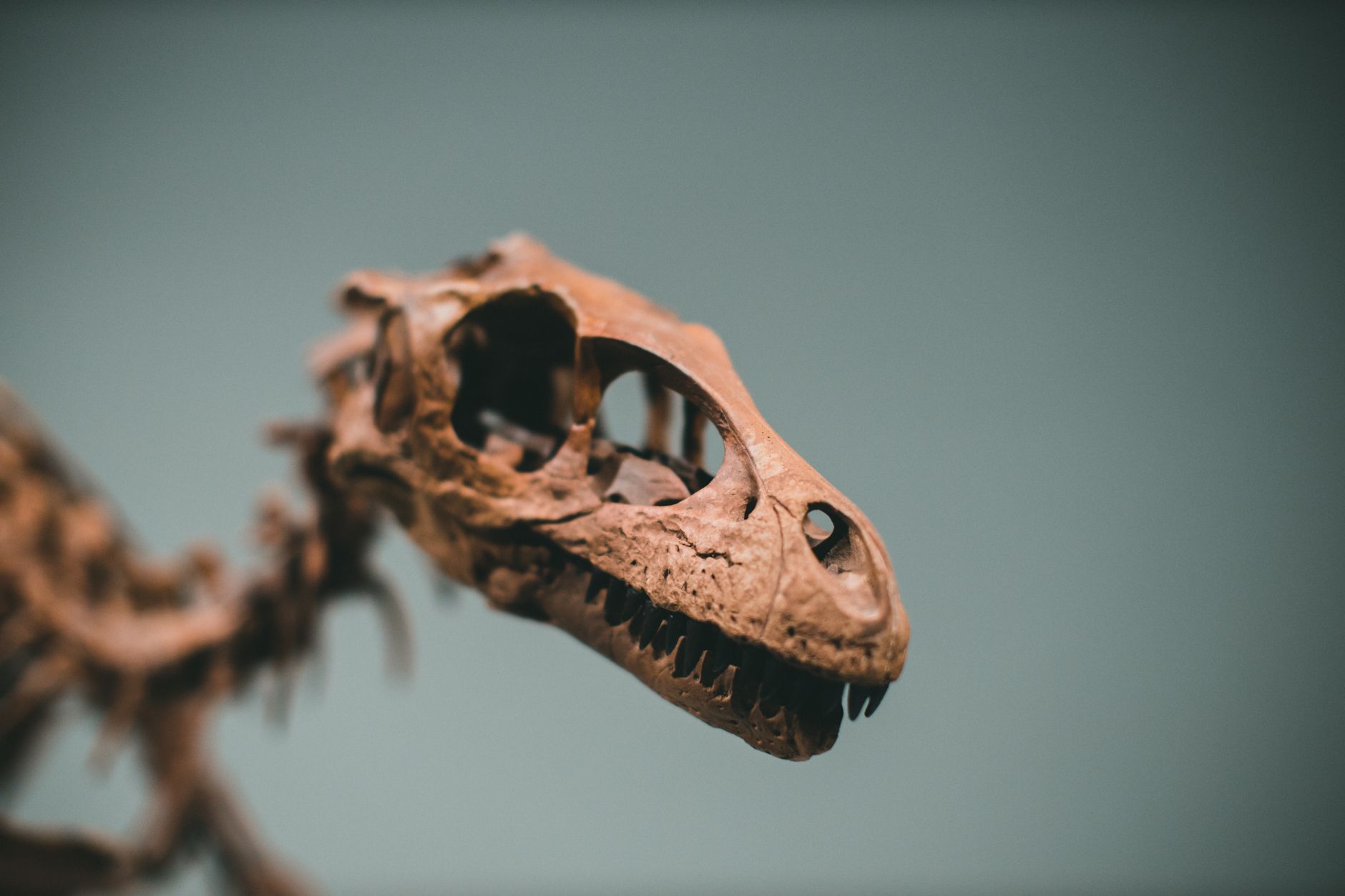
Aratasaurus museunacionali was a small, two-legged, carnivorous theropod dinosaur that lived during the Early Cretaceous period, approximately 115 million years ago. It is estimated to have been about 10 feet (3 meters) long.
How did the blood cells survive for 115 million years?
The preservation was likely due to a unique combination of factors: rapid burial in a low-oxygen environment, the presence of microbial mats, and early mineralization by a manganese oxide mineral called hausmannite. This mineral process effectively created a “mineralized record” of the delicate biological structures.
Why is there a debate about the discovery?
The debate is primarily over the shape of the structures. While the researchers found chemical evidence of blood (heme), some paleontologists, such as Mary Schweitzer, argue that the physical shape of the structures does not match the “nuclear bulge” expected in the red blood cells of reptiles and birds.
What is heme?
Heme is an iron-containing, ring-shaped molecule that is a vital component of hemoglobin. Its primary function is to facilitate the transport of oxygen within the blood cells of animals.
Scientists are now looking toward the future of paleontology, where the study of life’s history may rely as much on molecular chemistry as it does on the study of bone and tooth
References
Featured image: Image via Live Science