For generations, expectant and new mothers have swapped stories about “mommy brain” or “pregnancy brain.” This often-joked-about phenomenon describes a feeling of fogginess, forgetfulness, or increased absent-mindedness during and after pregnancy. But is it real? And if so, what physically happens to the brain during this incredible time of transformation?
Despite how common this experience is, scientific research into the actual physical changes the brain undergoes during pregnancy has been surprisingly limited. The biological shifts required to grow a new human are immense, impacting nearly every system in the body. Yet, the organ responsible for processing all this, the brain, remained largely a scientific “black box.”
Unpacking the “Mommy Brain” Phenomenon
The anecdotal evidence for “mommy brain” is strong. Many individuals report difficulty concentrating, misplaced items, or simply feeling less sharp than before pregnancy. For a long time, the scientific community lacked clear answers about the root cause. Was it just sleep deprivation? Stress? Hormones?
Traditional studies often compared brains before and after pregnancy, missing the dynamic changes happening throughout the nine months and the crucial postpartum period. This left a significant gap in our understanding.
A Groundbreaking Self-Study: Scanning the Neuroscientist’s Brain
Inspired to close this gap, neuroscientist Liz Chrastil embarked on a truly unique research project. Leveraging her expertise in learning, memory, and MRI technology, she decided to study the ultimate subject: herself.
The Unprecedented Methodology
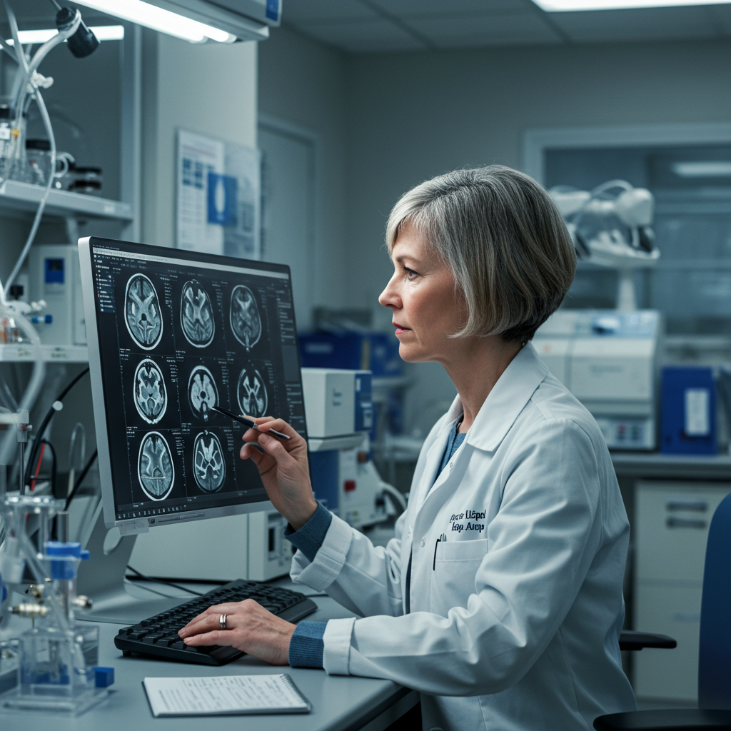
Timed with her journey through in vitro fertilization (IVF), Chrastil committed to an extraordinary protocol. She underwent a staggering 26 MRI scans of her own brain. These scans took place at multiple points: before conception, throughout her pregnancy, and for two full years into the postpartum period.
Each scanning session lasted around 40 minutes. This added up to over 1,000 minutes inside the MRI machine. Alongside the scans, she also provided corresponding blood samples to track hormone levels.
Chrastil was already very familiar with MRI technology. She knew it uses magnetic fields and radio waves, not radiation. Major health organizations like the American College of Obstetricians and Gynecologists (ACOG) consider it safe during pregnancy. She felt comfortable with the procedure, taking only minor comfort measures like adding foam to soften the sound.
Why Study the Pregnant Brain?
Chrastil’s motivation was clear. While anecdotal evidence of “mommy brain” was widespread, hard data on structural brain changes was scarce. Researchers like Laura Pritschet and Emily Jacobs, who collaborated on the later analysis of this data, recognized the need for a detailed, longitudinal look at the brain during pregnancy. Their goal was to map neuroanatomical changes and see how they correlated with fluctuating hormone concentrations, providing unprecedented insight into this understudied period.
Surprising Discoveries: Gray Matter Shrinkage and White Matter Shifts
The results of this unprecedented self-study, later published in the prestigious journal Nature Neuroscience, were eye-opening. They provided the first detailed map of how the human brain changes across the entire pregnancy and postpartum timeline.
What the Scans Revealed
One of the most significant findings involved gray matter. This is the brain tissue primarily responsible for vital functions like processing information, memory, sensory perception, language, and decision-making. The scans showed a notable decrease in the volume of gray matter. This reduction amounted to about 4%. The decrease wasn’t sudden; it occurred steadily throughout pregnancy, starting surprisingly early in the first few weeks. This change was widespread, affecting approximately 80% of brain regions. The reduction also correlated with rising levels of sex hormones, specifically estradiol and progesterone.
But that wasn’t the only change. The study also looked at white matter. This tissue forms the connections between different brain regions, acting like superhighways for information transfer. Here, the changes were different and more transient. The scans indicated that the structural integrity of white matter actually improved. This increase in white matter strength peaked around the second trimester of pregnancy. However, unlike gray matter, the white matter integrity returned to near its baseline level after the baby was born.
Is it Permanent?
While the white matter changes appeared temporary, the gray matter reduction seems more lasting. The study found that while there was a slight recovery in gray matter volume postpartum, it did not return to its pre-pregnancy levels within the two-year study period. Other research cited by the collaborators has shown that the “trace” of pregnancy brain changes can be detectable in neuroimaging data even decades later. This suggests the structural transformations are permanent etchings on the brain.
Despite these physical changes, Chrastil personally reported no noticeable difference in her cognitive abilities or how she felt she was thinking throughout the process.
Interpreting the Changes: Adaptive Wiring or Resource Trade-off?
The big question scientists face now is: what do these changes mean? The functional implications of the gray and white matter alterations observed in the study are not yet fully understood.
One prevailing theory, supported by some researchers, is that the gray matter reduction might be adaptive. This process is potentially similar to the “pruning” that occurs in adolescent brains. During adolescence, the brain refines neural connections, getting rid of weaker ones to make processing more efficient. In pregnancy, this pruning might serve to enhance specific brain regions involved in social cognition. The idea is that the pregnant brain might be rewiring itself to become more attuned to the needs of an infant, enhancing skills like recognizing cries or understanding non-verbal cues.
Another possibility is that the changes represent a trade-off. The immense physiological demands of pregnancy require vast resources. Perhaps some brain tissue volume decreases because resources are being diverted to support other critical processes in the body, like fetal development. The truth might be a combination of both adaptive wiring and resource allocation shifts.
Chrastil herself emphasizes the current lack of knowledge in this area. It is, she notes, “shocking how little scientists know” about brain changes during pregnancy. The research, at this stage, raises more questions than it currently answers.
Bridging the Knowledge Gap: Future Research and Perinatal Mental Health
This foundational self-study, though based on a single participant, served as a crucial proof-of-concept. It demonstrated that scanning throughout pregnancy is not only feasible but essential to capture the dynamic nature of these brain changes. The researchers have made their data openly available, encouraging other scientists to build upon these initial findings.
Why This Research Matters
Understanding the typical pattern of brain changes during pregnancy is vital. It establishes a baseline against which researchers can compare variations. This could have significant clinical implications, particularly for perinatal mental health.
Looking Ahead: Addressing Perinatal Mental Health
Perinatal mental disorders, such as postpartum depression, affect a substantial number of people who give birth – estimates suggest up to one in eight. The hope is that by mapping typical gray and white matter changes, researchers can identify deviations that might serve as early indicators or biomarkers for increased risk of these conditions.
Chrastil and her collaborators are already planning follow-up research. Their next step is a larger study involving 10 to 15 pregnant women. The long-term goal is to eventually scan hundreds of women, using fewer scanning points per individual to make it more scalable. This larger body of data could help confirm the initial findings and, critically, investigate the connection between observed brain changes and the risk trajectory of postpartum depression throughout pregnancy and into the postpartum period. The aim is to pinpoint opportunities for better prevention and intervention strategies.
Advocating for Inclusion: Pregnant Individuals in Scientific Research
This research also highlights a broader issue in scientific study: the historical underrepresentation and often outright exclusion of women and pregnant individuals from research, particularly imaging studies like MRIs. Despite evidence supporting the safety of MRI during pregnancy, concerns have often been used as a reason to exclude this population.
Researchers like Jacobs argue that this exclusion isn’t always strictly based on proven risk. Instead, it can reflect a systemic neglect of women’s unique physiological states and health issues within biomedical science. Actively advocating for the inclusion of pregnant participants in imaging studies is crucial. Chrastil’s protocol serves as a model, potentially encouraging other imaging centers to follow suit and help jumpstart this vital field of maternal brain research.
Expanding scientific understanding to include pregnancy, alongside other female-specific phenomena like menopause and hormone therapy, is essential. It’s a necessary step towards creating a more comprehensive, equitable, and ultimately beneficial science for everyone.
Frequently Asked Questions
What are the main physical changes observed in the brain during pregnancy?
Research, including a unique self-study by a neuroscientist, shows significant physical changes in the brain during pregnancy. The primary change is a decrease in the volume of gray matter, the tissue responsible for cognitive processing, memory, and decision-making. This reduction is about 4% and appears to be long-lasting. Changes also occur in white matter, the brain’s connections, which temporarily improve in structural integrity during pregnancy before returning to baseline postpartum.
Does pregnancy brain permanently affect cognitive abilities like memory?
While the physical gray matter reduction appears permanent, the impact on cognitive abilities like memory is unclear and may not be negative. Anecdotally, many report “mommy brain” fogginess. However, the gray matter changes are theorized to be an adaptive “pruning” process, potentially enhancing brain regions for social cognition and infant care. The neuroscientist who scanned her own brain felt no subjective difference in her thinking despite the observed physical changes. More research is needed to fully understand the functional outcome.
How might this research help with issues like postpartum depression?
Understanding the typical pattern of brain changes during pregnancy is a crucial first step in addressing perinatal mental disorders like postpartum depression. By mapping these standard changes, researchers hope to identify deviations or variations in brain structure or hormone correlation that might indicate a higher risk for depression or other mood disorders. This could potentially lead to the development of biomarkers for early identification and allow for better prevention and intervention strategies for affected individuals.
—
This groundbreaking study represents a vital first step. It moves beyond anecdote to provide concrete evidence of the profound structural changes the brain undergoes during pregnancy. While the meaning of these changes is still a frontier of scientific discovery, the research opens exciting avenues for understanding the complex interplay of hormones, environment, and brain plasticity during this unique life stage, with significant implications for maternal health and well-being.


