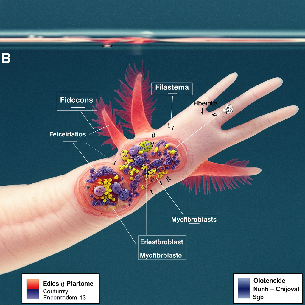
Axolotls (Ambystoma mexicanum) possess an extraordinary ability to regenerate complex body parts, including full limbs, hearts, and even parts of their brains. This remarkable feat involves a sophisticated process where cells at the injury site form a mass called a blastema, which then rebuilds the missing structure with astonishing accuracy. A critical aspect of this process is ensuring the regenerating limb forms the correct segments along the shoulder-to-fingertip axis, known as the proximodistal (PD) axis. But how does the blastema know exactly what segment to regenerate?
The Puzzle of Positional Identity
When an axolotl limb is amputated, the remaining tissue “remembers” its original position along the PD axis. Cells in the blastema receive this positional information, allowing them to reconstruct only the missing parts – for example, an amputation below the elbow regenerates only the forearm and hand, not a new upper arm. This “positional identity” is genetically determined by patterning genes like Hox and Meis, which establish unique molecular signatures along the limb axis. While the genetic basis is understood, the precise mechanism by which this continuous positional information is established and interpreted during regeneration has remained a key question.
Retinoic Acid: A Master Regulator
Retinoic acid (RA), a molecule derived from Vitamin A, is a well-known signaling molecule crucial for development in many animals, including the initial formation and patterning of limbs. In both developing and regenerating limbs, RA is understood to play a significant role in specifying proximal identity (closer to the body). Studies have shown that proximal blastemas (closer to the shoulder) naturally have higher levels of RA signaling compared to distal blastemas (closer to the fingertips). Furthermore, artificially increasing RA in distal blastemas can reprogram cells to think they are more proximal, leading to the regeneration of duplicated proximal structures like extra forearms or upper arms.
While the link between RA and proximal identity is clear, how the distinct RA signaling levels are established and maintained between proximal and distal blastemas during regeneration wasn’t fully understood.
The Breakthrough: The Role of RA Breakdown
New research sheds light on this mystery, pointing to the controlled breakdown of RA as the critical factor determining positional identity. The study focused on the enzyme CYP26B1, which is known to degrade RA.
Researchers examined the expression of genes involved in RA synthesis, signaling, and degradation in blastemas formed at different positions along the PD axis. While RA synthesis enzymes didn’t show clear patterns explaining the RA gradient, the RA degradation enzyme Cyp26b1 did. Cyp26b1 expression was found to be significantly higher in distal blastemas, particularly in the mesenchymal cells that form the bulk of the regenerating tissue, and decreased progressively towards proximal blastemas. This spatial pattern is the inverse of the known RA activity gradient (high proximally, low distally), strongly suggesting that higher RA breakdown in distal regions, rather than differential synthesis, is responsible for keeping RA levels low there.
Experimental Proof: Blocking Breakdown Reprograms Regeneration
To directly test the role of CYP26B1, scientists used a pharmacological inhibitor (Talarozole) to block its activity in regenerating distal blastemas. The results were striking:
Inhibiting CYP26B1 significantly increased RA signaling in distal blastemas.
This increase in RA signaling mimicked the effects of adding excess RA, causing the distal blastemas to regenerate duplicated proximal skeletal elements (forearms or upper arms), depending on the inhibitor concentration.
This reprogramming involved downregulating distal patterning genes (Hoxa13) and upregulating proximal genes (Meis1, Meis2).
This experiment provided clear functional evidence that CYP26B1-mediated RA breakdown is essential for maintaining the correct low RA levels distally, preventing the misidentification of distal segments as proximal.
Identifying Key Downstream Players: The Importance of Shox
The RA gradient established by CYP26B1 breakdown influences the expression of RA-responsive genes that ultimately build the limb segments. The study identified Shox (Short stature homeobox) as a key gene that is both responsive to RA and expressed differently along the PD axis (higher proximally).
To understand Shox‘s specific role, researchers used CRISPR/Cas9 gene editing to eliminate Shox function in axolotls. Shox knockout animals showed a remarkable and specific defect: their proximal limb segments (upper arm and forearm) were significantly shorter than normal, while their distal segments (hand) developed correctly and had normal length. Further analysis revealed that the cartilage structures in the proximal segments of Shox knockouts failed to properly undergo endochondral ossification – the process where cartilage is replaced by bone necessary for bone lengthening and maturation.
Crucially, Shox knockout axolotls were still perfectly capable of regenerating complete limbs, although the regenerated proximal segments still exhibited the characteristic shortening and ossification defects. This indicates that while Shox is not required for the fundamental regeneration process itself, it is essential for the proper growth and skeletal maturation of the proximal limb segments based on their determined identity.
A New Model for Axolotl Limb Patterning
This research proposes a refined model for how axolotls achieve precise limb regeneration:
- Positional Memory: Cells in the blastema retain positional information from the stump.
- RA Gradient Establishment: High expression of the RA-degrading enzyme CYP26B1 in distal blastema cells (and low expression proximally) creates a gradient of Retinoic Acid (RA) activity along the PD axis (high proximally, low distally).
- Differential Gene Activation: This RA gradient activates or represses RA-responsive genes based on their sensitivity and the local RA concentration.
- Segment Specification: High RA levels in proximal blastemas activate genes like Shox and Meis1, specifying proximal identity and driving processes like endochondral ossification necessary for proper proximal segment formation and growth. Low RA levels in distal blastemas allow for the expression of distal genes like Hoxa13* and the formation of distal structures through Shox-independent mechanisms.
In essence, the axolotl’s ability to regenerate the correct limb segment isn’t just about having the right building blocks; it’s about tightly controlling the levels of key signaling molecules like Retinoic Acid through enzymes like CYP26B1, which in turn orchestrate the precise genetic programs required for patterning and building each unique segment along the limb. This fundamental insight into regenerative biology could have implications for future strategies in regenerative medicine.


