
Scientists have made a groundbreaking discovery within the complex world of human cells. They’ve identified a previously unknown structure, now named the “hemifusome.” This novel organelle is believed to play a crucial role in cellular recycling and the internal sorting of materials. The finding challenges long-held assumptions about how cells manage their internal components. Researchers from the University of Virginia (UVA) School of Medicine and the National Institutes of Health (NIH) led this exciting work. Their study, published in Nature Communications, suggests a completely new pathway for these vital cellular functions.
Understanding how cells sort and process materials is fundamental to life. These processes are often disrupted in various diseases. The discovery of the hemifusome opens new avenues for research. It could potentially lead to better insights into conditions ranging from Alzheimer’s disease to viral infections and certain cancers.
Unveiling the Hemifusome: A New Cellular Player
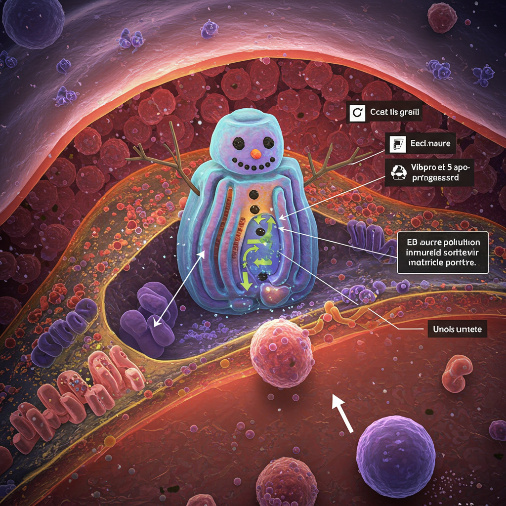
What exactly is this newly found structure? Researchers describe the hemifusome not as static, but as a temporary formation. It consists of two vesicles, which are small, membrane-bound sacs used for transport within the cell. These two vesicles are joined by a unique partial membrane connection. Scientists call this connection a hemifusion diaphragm.
Unlike full fusion, this partial state allows the vesicles to interact while maintaining separate identities. This configuration is a key characteristic. The hemifusome has a distinctive shape, resembling a “snowman wearing a scarf,” according to one description. This involves a smaller vesicle attached to a larger one. A thin border, the hemifusion diaphragm, lies between them.
How This ‘Snowman’ Organelle Was Discovered
Finding the hemifusome was a significant challenge. Traditional microscopy techniques often require harsh processing steps. These steps can alter or destroy delicate cellular structures. The hemifusome is also ephemeral. It forms only when needed and disappears quickly. This fleeting existence makes real-time observation difficult.
The breakthrough came with advanced imaging technology. Researchers utilized cryo-electron tomography (cryo-ET). This technique rapidly freezes cells. It preserves their internal structures in a near-native state. Cryo-ET provides incredibly detailed 3D snapshots. These images have nanometer resolution. They revealed structures previously invisible. The team, including co-leader Dr. Seham Ebrahim of UVA, used cryo-ET available at UVA’s Molecular Electron Microscopy Core. This technology was essential for visualizing the transient hemifusome.
The study examined four different mammalian cell types: COS-7, HeLa, RAT-1, and NIH/3T3. To their surprise, hemifusomes were not rare. They were consistently observed at the outer edges of these cells. Nearly 1 in 10 vesicles found in these regions were identified as hemifusomes. This suggests they are common components rather than unusual anomalies.
Decoding the Hemifusome’s Potential Role
The primary suspected function of the hemifusome is in cellular recycling. Specifically, it appears involved in forming multivesicular bodies (MVBs). MVBs are crucial structures inside cells. Think of them as recycling centers or sorting hubs. They are responsible for sorting and removing unwanted proteins. MVBs are vital for numerous cellular functions. These include immune responses and intercellular communication. They also help protect against neurological diseases.
Prior scientific understanding largely credited MVB formation to a protein-based system. This system is known as the ESCRT (Endosomal Sorting Complexes Required for Transport) system. The hemifusome, however, seems to facilitate MVB formation differently. Researchers propose a mechanism based on lipid remodeling. This is distinct from protein scaffolding.
Observations using electron tomography support this. Hemifusomes exist in that unique, partially fused membrane state. They were consistently associated with tiny lipid-rich droplets. These are called proteolipid nanodroplets (PNDs). These PNDs are typically found lodged at the rim of the hemifusion site. Scientists propose PNDs play a vital role. They may act as anchors or scaffolds. They could guide the formation and stability of the hemifusome. They might even initiate the de novo (from scratch) assembly of the smaller vesicle within the hemifusome structure. This proposed mechanism of de novo vesiculogenesis is different from classic vesicle fusion processes.
A New Pathway for Cellular Housekeeping
The discovery of the hemifusome suggests a novel pathway. It challenges the previous understanding of vesicle formation. This new pathway relies on structural and biophysical cues from PNDs. This contrasts with complex protein machinery like the ESCRT system. It also differs from mechanisms relying on large lipid donations.
Further evidence supporting this new pathway came from experiments using gold nanoparticles. These particles were used as markers for endocytic activity. They entered known endosomes and lysosomes. However, they were absent from hemifusomes. This suggests hemifusomes operate independently of the classical endocytic pathway. They seem separate from typical cargo sorting mechanisms involving proteins like ESCRT.
The study also observed complex formations. These were called compound hemifusomes. They contained multiple partially fused vesicles. These formations are suggested as potential precursors to MVBs. The researchers propose an alternative model for MVB formation. In this model, vesicles bud inward via hemifusion. This budding is guided by PNDs. This is a significant contrast to the canonical ESCRT-driven inward budding mechanism.
Implications for Health and Disease
The implications of finding the hemifusome are considerable. This new organelle sheds light on a novel method cells use to sort and move materials internally. Such insights are critical for understanding diseases where these processes fail.
Conditions like Alzheimer’s disease involve the buildup of misfolded proteins. Many viral infections hijack cellular machinery. Certain cancers disrupt cellular waste disposal. Understanding the hemifusome’s role could provide valuable insights into these conditions.
Genetic disorders offer another key area of impact. Many genetic diseases are rooted in problems with cellular cargo handling. Hermansky-Pudlak syndrome is one example. This rare genetic disorder affects albinism, vision, lungs, and blood clotting. These issues are linked to defects in cellular cargo management. Studying hemifusomes could illuminate the disruptions seen in such diseases. It might potentially lead to new therapeutic strategies.
Future Research Directions
This discovery is an initial, exciting step. Scientists are eager to learn more about the hemifusome. Future work needs to identify the specific proteins involved in its structure and function. Researchers also need to understand how PNDs are created and regulated. Exploring the presence and function of hemifusomes in other cellular locations is also critical.
The study highlights the power of advanced imaging techniques. Cryo-ET revealed previously hidden cellular structures. This suggests there may still be much to uncover within cells. Once their role is fully confirmed, hemifusomes could be recognized as a new class of intermediate structures involved in fusion processes in mammalian cells.
Frequently Asked Questions
What is the hemifusome and what does it do inside cells?
The hemifusome is a newly discovered organelle found in human cells. It is a temporary structure formed by two vesicles (small sacs) connected by a partial membrane barrier. Scientists believe its main role is in cellular recycling and sorting cargo, specifically suggesting a new way that cells form multivesicular bodies (MVBs), which are internal recycling centers.
How was the hemifusome discovered if it’s only temporary?
Discovering the hemifusome required advanced technology because it is temporary and difficult to see with traditional methods. Researchers used cryo-electron tomography (cryo-ET). This technique flash-freezes cells, preserving their delicate internal structures in a near-native state and allowing for detailed 3D imaging that revealed the hemifusome’s unique shape and structure.
Why is the hemifusome discovery important for understanding health and disease?
The discovery is significant because it reveals a potentially new pathway for cellular recycling and sorting, processes vital for cell health. When these processes fail, it can contribute to diseases like Alzheimer’s, viral infections, certain cancers, and genetic disorders like Hermansky-Pudlak syndrome. Understanding the hemifusome could offer new insights into how these diseases develop and potentially lead to new therapeutic targets.
This breakthrough expands our understanding of the intricate machinery within human cells. The hemifusome represents a new frontier in cell biology research. Future studies are expected to shed more light on its precise functions and potential impact on human health, potentially revolutionizing how we approach diseases linked to cellular cargo management. The journey to fully understand this ‘snowman’ shaped organelle has just begun.