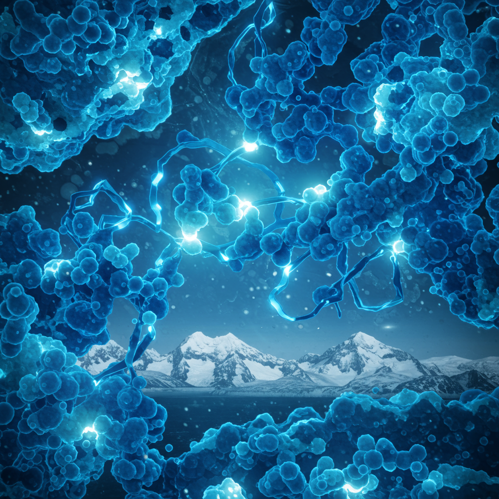
Imagine exploring Earth’s most frigid landscapes – towering glaciers, high mountain peaks, or the depths of permanently frozen ground water. While incredibly beautiful, these icy domains also harbor microscopic life with extraordinary adaptations. Among them are unusual molecules poised to revolutionize fields like neuroscience: rare blue proteins called cryorhodopsins, discovered in cold-loving microbes. These unique proteins show remarkable potential as prototypes for molecular on-off switches, offering precise control over cellular activity using light.
Discovery in the Deep Chill: Uncovering Cryorhodopsins
Structural biologist Kirill Kovalev embarked on a quest to find unusual molecules within these cold environments. Driven by a passion for solving biological puzzles, Kovalev focused on rhodopsins. These are a diverse family of colorful proteins used by many microorganisms, particularly in aquatic settings, to capture sunlight for energy. Some rhodopsins have already been modified for use in optogenetics. This powerful technique allows neuroscientists to control the electrical firing of neurons simply by shining light on them, acting like a light-activated switch. Kovalev sought rhodopsins with novel abilities, perhaps even enabling light control over chemical reactions.
His extensive study of rhodopsins led him to a surprising observation while browsing online protein databases. He noticed a peculiar structural feature shared exclusively by microbial rhodopsins originating from extremely cold locations, like glaciers and high mountain ranges. This was unexpected, as rhodopsins are more commonly associated with seas and lakes. The remarkable similarity among these cold-climate rhodopsins, despite evolving in distant geographical areas, strongly suggested they played a vital role in surviving frigid conditions. Recognizing their unique adaptation to the cold, Kovalev aptly named this new group “cryorhodopsins.”
The Mystery of Color: Why Blue Matters
The natural next step was to understand these cryorhodopsins more deeply: their structure, function, and, crucially, their color. A rhodopsin’s color is fundamental to its function, determining which wavelengths of light it absorbs and reflects. Most known rhodopsins appear pink-orange because they absorb green and blue light, which triggers their activation. Scientists are actively seeking a diverse palette of rhodopsin colors. This diversity allows for more precise and complex control over neuronal activity using different light wavelengths.
Blue rhodopsins, in particular, have been highly sought after. Why? Because they are activated by red light. Red light offers a significant advantage: it penetrates biological tissues far more deeply and less invasively than shorter wavelengths like green or blue. This makes blue-light-activated (red-light-responsive) rhodopsins ideal candidates for applications deep within the body, such as controlling brain cells or other embedded tissues without complex surgery.
Kovalev and his collaborators were amazed to find that the cryorhodopsins they examined in the lab exhibited a surprising array of colors. Most importantly, some were distinctly blue. Their findings, detailing the characterization of this novel group of microbial rhodopsins, were published in the journal Science Advances.
Decoding the Structure Behind the Blue
A protein’s color is directly linked to its molecular structure. Small changes in the arrangement of atoms can shift the wavelengths of light the protein absorbs and reflects, thereby changing its observed color. As Kovalev noted, “I can actually tell what’s going on with cryorhodopsin simply by looking at its color.”
Using advanced structural biology techniques, including X-ray crystallography and cryo-electron microscopy (cryo-EM), Kovalev and his team pinpointed the secret behind the cryorhodopsins’ blue hue. The blue color is conferred by the very same rare structural feature that Kovalev initially spotted in the protein databases. This specific structural motif dictates how the protein interacts with light, causing it to absorb red light and appear blue. Understanding this fundamental structural basis for the blue color is a critical breakthrough. “Now that we understand what makes them blue,” Kovalev explained, “we can design synthetic blue rhodopsins tailored to different applications.” This knowledge opens the door to engineering customized molecular tools.
Beyond Color: Bidirectional Switching Potential
The potential of cryorhodopsins extends beyond their unique color. Experiments conducted by Kovalev’s collaborators on cultured brain cells demonstrated a remarkable ability. When cells engineered to express cryorhodopsins were exposed to UV light, it triggered the flow of electric currents within them. This confirmed their light sensitivity and potential to influence cellular electrical states.
Even more intriguing was their response to subsequent light pulses. If researchers followed the initial UV exposure with green light, the cells became more excitable – effectively switching their activity “on.” Conversely, if they used UV followed by red light, the cells’ excitability decreased, demonstrating an “off” switch effect. This bidirectional control – the ability to both increase and decrease cellular electrical activity with different light signals – is a highly valuable property for molecular switches.
Tobias Moser, a Group Leader involved in the study, highlighted the significance: “New optogenetic tools to efficiently switch the cell’s electric activity both ‘on’ and ‘off’ would be incredibly useful in research, biotechnology and medicine.” He cited their ongoing work developing optical cochlear implants to restore hearing, where precise light-based control of neurons is essential. While cryorhodopsins aren’t yet ready for clinical use, Kovalev considers them an excellent prototype. “They have all the key features that, based on our findings, could be engineered to become more effective for optogenetics,” he stated.
A Deeper Mystery: UV Sensing in Cold Extremes
Investigating the natural role of cryorhodopsins in their native habitats revealed another fascinating property. Collaborators using advanced spectroscopy techniques showed that cryorhodopsins are uniquely slow in their response to light compared to other rhodopsins. Furthermore, they demonstrated the ability to sense UV light, even in the weak sunlight of places like Hamburg during winter. This suggested a potential function as photosensors, enabling the microbes to “see” UV light – a capability previously unknown among other cryorhodopsins.
Why would cold-adapted microbes need to sense UV light? Kovalev pondered whether this might be linked to the harsh conditions of their environment. High-altitude and high-latitude cold regions, like mountain peaks and glaciers, often experience intense UV radiation that can damage biological molecules. The hypothesis emerged that cryorhodopsins might function as UV sensors, alerting microbes to harmful radiation so they can initiate protective measures.
Adding weight to this idea, Kovalev and collaborators discovered that the gene encoding cryorhodopsin is consistently found alongside a gene for a tiny protein of unknown function. This strong genetic linkage suggested they were inherited together and likely function as a pair. Could this small protein be a messenger, relaying the UV signal from the cryorhodopsin sensor on the cell membrane into the cell’s interior?
Using the powerful AI tool AlphaFold, the team predicted that five copies of this small protein could assemble into a ring structure and interact directly with the cryorhodopsin. Their model suggests the small protein sits ready near the cryorhodopsin inside the cell. Upon detecting UV light, cryorhodopsin might signal the small protein, causing it to detach and carry the “UV detected” message to other parts of the cell. This potential new mechanism for light-sensitive signal transmission is a significant discovery.
Technical Hurdles and Future Horizons
Unlocking the secrets of cryorhodopsins presented significant technical challenges. These proteins are structurally very similar, meaning even subtle atomic shifts can drastically alter their properties. Studying them required going beyond standard methods. Kovalev employed a cutting-edge 4D structural biology approach. This method combines high-resolution techniques like X-ray crystallography at facilities like EMBL Hamburg’s P14 beamline and cryo-EM with the dynamic element of protein activation by light. The unique setup at EMBL was crucial for this work.
Another hurdle was the extreme light sensitivity of the cryorhodopsins themselves. Researchers had to learn to handle samples and conduct experiments in near-complete darkness to prevent unintended activation.
Discovering such unique molecules highlights the value of scientific expeditions to remote locations and studying how organisms adapt to extreme environments. These studies provide insights into novel biological mechanisms and yield promising prototypes for new tools in science, technology, and medicine. Cryorhodopsins, with their rare blue color, bidirectional switching capability, and potential UV sensing role, exemplify this potential. They offer an exciting blueprint for designing next-generation light-controlled molecular tools that could benefit neuroscience, biotechnology, and perhaps future medical treatments.
Frequently Asked Questions
How do cryorhodopsins from cold microbes act as molecular switches?
Cryorhodopsins are light-sensitive proteins found in cold environments. Researchers discovered they can control cellular electrical activity when exposed to specific light wavelengths. UV light can induce electric currents. Following UV, green light increases cell excitability (“on” switch effect), while UV/red light reduces it (“off” switch effect). This ability to bidirectionally control electrical states makes them potential prototypes for light-activated molecular switches in cells.
Which specific techniques and collaborations were key to discovering cryorhodopsins?
The discovery involved multiple advanced techniques and international collaboration. Structural biologist Kirill Kovalev initially spotted them in protein databases. Researchers used advanced structural biology methods like X-ray crystallography (at EMBL Hamburg) and cryo-electron microscopy. Spectroscopy revealed their light response, and the AI tool AlphaFold was used to predict the interaction with a potential messenger protein. Collaborators were involved from EMBL, University Medical Center Göttingen, Goethe University Frankfurt, and institutions in Spain and the Netherlands.
What potential applications do cryorhodopsins hold for biotechnology and medicine?
Cryorhodopsins are seen as promising prototypes for new optogenetic tools. Their ability to control cellular electrical activity with light could be invaluable in neuroscience research, allowing precise manipulation of neurons. In medicine, potential future uses include developing optical cochlear implants to restore hearing, where light-based neuronal control is needed. Understanding their structure also allows designing synthetic versions for tailored applications in biotechnology and medicine.
Paving the Way for New Light-Activated Tools
The discovery and characterization of cryorhodopsins represent a significant step forward in understanding microbial life in extreme environments and in developing advanced molecular tools. These rare blue proteins, isolated from organisms thriving in intense cold, possess unique properties that make them ideal starting points for engineering new optogenetic switches. Their potential for precise, light-based control over cellular activity, coupled with the insights gained into their structure and function, offers exciting possibilities for future research, technological innovation, and potential therapeutic applications in fields like neuroscience and beyond. Further research will focus on optimizing these fascinating molecules for practical use, building upon the fundamental knowledge uncovered by this pioneering work.
Word Count Check: 1150


