Human bipedalism, our unique ability to walk upright on two legs, fundamentally defines our species. This distinctive form of locomotion is made possible by a remarkably shaped pelvis, vastly different from our ape relatives. For centuries, scientists have pondered the precise developmental and genetic changes that sculpted the human pelvis, particularly the ilium, to support such a pivotal evolutionary leap. A groundbreaking study, published in Nature, now unveils the intricate genetic and mechanistic shifts in embryonic development that paved the way for human upright walking and successful childbirth.
The Pelvic Blueprint: A Human-Defining Adaptation
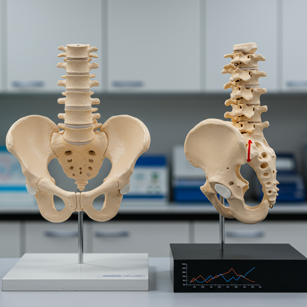
Our familiar bowl-shaped pelvis, with its short, wide iliac blades curving along the sides of the body, is an anatomical marvel. It provides critical stability for walking and running, supports internal organs, and facilitates the birth of large-brained, broad-shouldered babies. Compared to the tall, narrow ilia of other primates like chimpanzees, this human pelvic structure represents a profound evolutionary novelty. The question of how this unique morphology developed has long remained a mystery, sparking much speculation since Darwin first linked upright locomotion to the human lineage. While bipedalism appeared early in hominin evolution, predating significant brain enlargement, the exact biological underpinnings of this giant step have been elusive.
Shift One: Reorienting Cartilage Growth
The research identifies the first major innovation as a perpendicular (transverse) reorientation of the iliac cartilage growth plate. Most bones, including the ilia in other primates and mice, develop from cartilage models where chondrocyte growth plates align longitudinally along the head-to-tail axis. However, the human ilium takes a dramatically different path.
Initially, at around gestational day 45 (E45), the human ilium model appears similar to other primates, aligned cranio-caudally. But by E53–E54, something extraordinary happens: chondrocyte proliferation and differentiation rapidly shift to an anterior-posterior direction. This reorientation culminates by E72, with the growth plate zones aligning horizontally, or transversely. This pivotal shift simultaneously widens the ilium while reducing its height, creating the characteristic broad human hip. Researchers were surprised by this immediate, combined effect, expecting a more gradual, step-wise progression.
Molecular Orchestra: Guiding Pelvic Shape
The study delved into the molecular mechanisms driving this reorientation. It revealed complex regulatory changes within an integrated chondrocyte–perichondral–osteoblast pathway. Key players include SOX9, a transcription factor crucial for chondrification, and its downstream effector, PTH1R, involved in chondrocyte proliferation. Spatially, SOX9 shows an asymmetric pattern, localizing strongly to the anterior and posterior ends, while PTH1R becomes restricted to the anterior side, marking a proliferation initiation site.
Clinical evidence supports their importance. Patients with conditions like Campomelic Dysplasia (due to SOX9 mutations) or Jansen’s Metaphyseal Chondrodysplasia and Eiken Syndrome (linked to PTH1R mutations) often exhibit narrow, abnormally ‘tall’ ilia, underscoring these genes’ role in achieving human-like transverse flaring. External signaling cues, such as pleiotrophin (PTN) and midkine (MK) from adjacent mesenchyme, and parathyroid hormone (PTH) from the perichondrium, also contribute to this intricate process. Another crucial gene, ZNF521, which interacts with PTH1R, was implicated in this network.
Further supporting an evolutionary basis, the study found strong overlaps between human accelerated regions (HARs)—segments of the human genome that have undergone rapid changes since our divergence from other primates—and regulatory elements in the mesoderm and early cartilage at the critical E53–E54 expansion stage. These HARs are significantly enriched for genes related to skeletal morphogenesis and cell differentiation, pointing to ancient positive selection pressures that shaped this unique growth plate reorganization.
Shift Two: A Delayed Dance of Bone Formation
The second major innovation in human pelvic development involves a delayed and unique pattern of ossification, or bone formation. In most primates and mice, primary ossification begins internally, at the mid-shaft of the ilium, rapidly invading the cartilage model. Humans, however, follow a distinct and significantly slower trajectory.
The human primary ossification center (POC) initiates around E57, not in the mid-shaft, but unilaterally near the posterior border, close to the sacrum. From this point, ossification extends anteriorly in a radial fashion, remarkably remaining restricted to the periphery (perichondrium) of the cartilaginous model. Crucially, internal ossification is substantially delayed, not beginning to deeply enter the ilium until around 24 weeks of gestation—up to 16 weeks later than in quadrupedal primates. Blood vessels, too, extend along the peripheral surface, contrasting with other species where they penetrate deep into the internal cartilage early on. This unique peripheral ossification, coupled with delayed internal hardening, allows the ilium to maintain its complex shape as it continues to grow.
Molecular Orchestration of Ossification
RNA velocity analysis revealed that both perichondral cells and fibroblasts contribute to this unique peripheral osteoblast population—a novel finding in human development. Gene regulatory networks play a vital role here, with RUNX2 identified as a master regulator in the early perichondrium. Later, FOXP1/FOXP2 emerge as top regulators in perichondral cells and osteoblasts. Transgenic mouse models demonstrated that a specific human RUNX2 enhancer showed activity in the iliac perichondria, an activity absent in the corresponding chimpanzee ortholog. This suggests a human-specific regulatory evolution.
The evolutionary signature for this ossification shift is also strong. Researchers identified 70 HARs within perichondral and osteoblast-specific regulatory elements, significantly enriched for terms like ‘developmental growth’ and ‘delayed bone ossification’. This indicates that this unique ossification pattern was shaped by complex polygenic selection and is under ongoing evolutionary constraint.
Interconnectedness: A Masterpiece of Evolution
These two developmental innovations—the transverse reorientation of the growth plate and the delayed peripheral ossification—are not isolated events but deeply interconnected. The transverse growth permits the asymmetrical widening of the ilium instead of merely increasing its height. This widening is sustained by an Anterior Growth Zone (AGZ), which remains cartilaginous for an extended period. This AGZ is crucial for the formation of key attachment points like the anterior superior and inferior iliac spines (ASIS/AIIS), which are vital for bipedal muscle function.
The soft tissue environment also plays a critical role. Muscles essential for bipedalism, such as the gluteus medius, gluteus minimus, and rectus femoris, along with the iliofemoral ligament, directly attach to the developing cartilaginous ilium and the AIIS early in embryonic development. Strong HAR enrichments were observed in muscle, perimysium, and tenocyte cell types during these critical stages, suggesting that early muscle anchoring and contractions, potentially through mechanosensitive RUNX2, influence the pelvic blade’s parasagittal orientation and the pace of the advancing peripheral ossification front.
A Three-Stage Evolutionary Model for Bipedalism
Based on these findings, the authors propose a three-step model for the evolutionary reconfiguration of the human pelvis:
- Early Adaptation (8–5 Million Years Ago): An abrupt or gradual shift occurred from vertical to transverse chondrocyte growth. This change supported increased lateral stabilization, crucial for early facultative bipedalism as hominins diverged from African apes. Fossil evidence from Sahelanthropus, Orrorin, and Ardipithecus hints at early upright postures during this period.
- Fixation and Refinement (5–2 Million Years Ago): As hominins transitioned from facultative to obligatory bipedalism, the transverse growth plate orientation became fixed. This stage saw concomitant shifts in ossification, likely beginning at the posterior ilium to allow for anterior growth, effectively responding to evolving muscle functions. The Australopithecus pelvis, famously seen in “Lucy,” showcases more developed bipedal traits like flaring hip blades.
- Advanced Bipedalism and Birth (2 Million Years Ago Onwards): More recently, ossification timing became significantly delayed. This delay enabled enhanced growth and the retention of the complex iliac shape, crucial for accommodating the demands of running and the birthing of increasingly large-brained, broad-shouldered babies—the “obstetrical dilemma.”
This new research compels a rethinking of fundamental assumptions in human evolution. It argues that all hominin fossils from the point of this mechanistic shift onward developed their pelvis uniquely, diverging from any other primate. Consequently, subsequent evolutionary changes, such as increases in human brain size, should be interpreted within this novel, human-specific pelvic growth model, rather than models based on chimpanzee or other primate development.
Frequently Asked Questions
What are the two main developmental shifts in the human pelvis for bipedalism?
The study identifies two pivotal shifts. First, a heterotopic (spatial) shift in the iliac cartilage growth plate orientation occurs. Unlike other primates, human embryonic cartilage cells in the ilium reorient from a longitudinal (head-to-toe) axis to a perpendicular (transverse or anterior-posterior) direction by gestational day 72, making the pelvis shorter and wider. Second, there’s a heterochronic (timing) and heterotopic (spatial) shift in ossification. Human iliac bone formation is delayed, initiating posteriorly and spreading radially along the periphery of the cartilage, with internal ossification occurring significantly later (up to 16 weeks after other primates), allowing for the unique pelvic shape to be maintained during growth.
Which genes or molecular pathways are crucial for these pelvic changes?
Several key molecular players are identified. The transverse cartilage reorientation is regulated by an integrated network involving SOX9 (critical for cartilage formation) and its downstream effector PTH1R (for chondrocyte proliferation), along with external signals like ZNF521. For the delayed ossification, transcription factors RUNX2 and FOXP1/FOXP2 are master regulators, guiding the unique peripheral bone formation. Evolutionary evidence from Human Accelerated Regions (HARs) overlapping regulatory elements for these genes underscores their significance in human-specific pelvic development.
How does this new understanding of human pelvis development challenge previous evolutionary theories?
This groundbreaking research suggests a “complete mechanistic shift” in human pelvic development, unique among primates from early embryonic stages. It indicates that the human pelvis evolved under a distinct developmental program. This challenges previous assumptions that might have extrapolated from chimpanzee or generalized primate developmental models. The findings imply that once this unique growth model emerged, all subsequent evolutionary pressures, including increases in fetal brain size and the “obstetrical dilemma,” acted upon an already fundamentally re-engineered pelvic structure, providing a fresh perspective on the interplay between bipedalism, childbirth, and human evolution.