
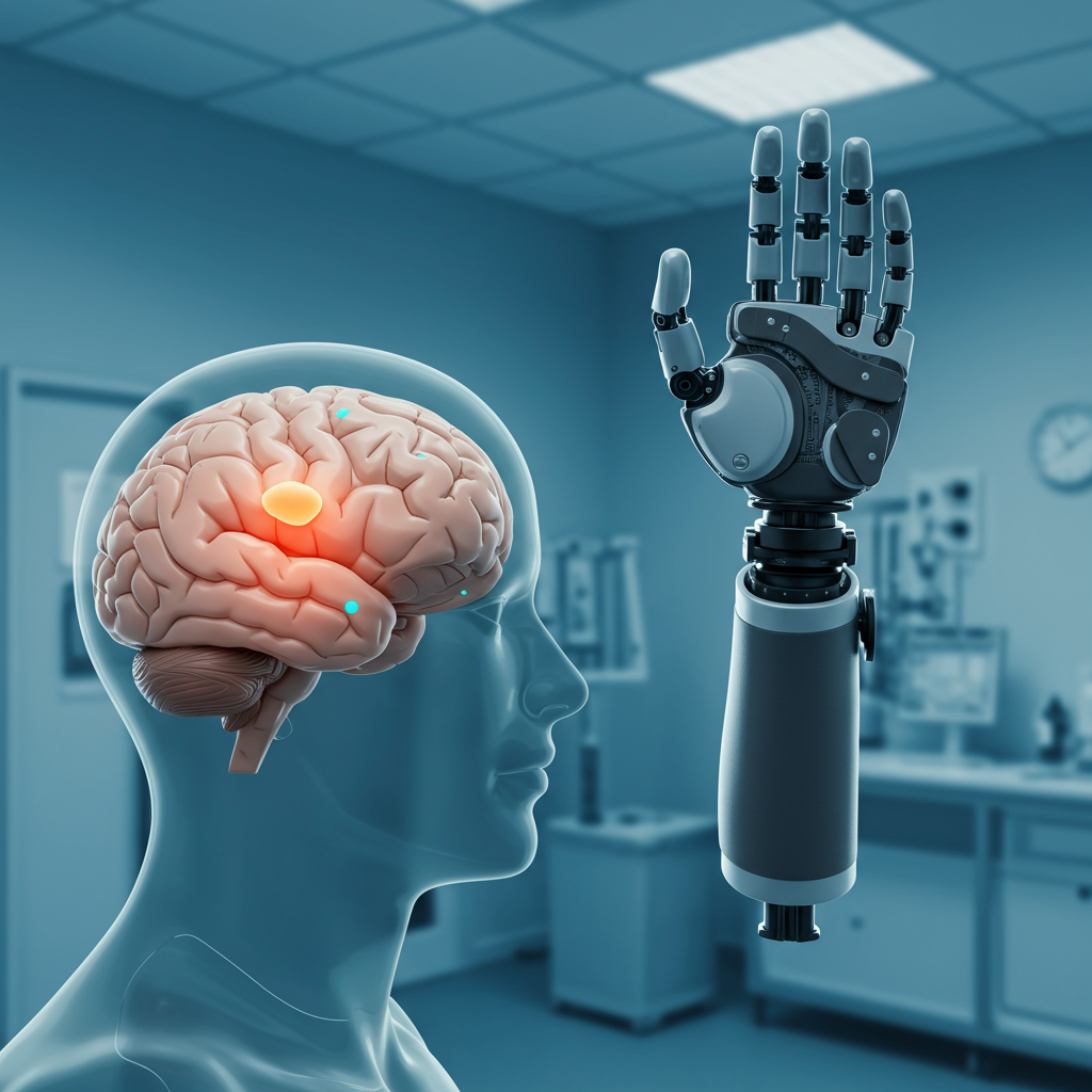
A groundbreaking study has unveiled a surprising truth about how the adult brain responds to limb loss, challenging a fundamental neuroscience tenet. Contrary to decades of belief, new research suggests that the brain’s body map for an amputated arm remains remarkably stable, refusing to be “rewired” or taken over by neighboring body parts. This revelation has profound implications for understanding phantom limb sensations, advancing prosthetics, and even redefining our understanding of adult brain plasticity.
The Enduring Mystery of Brain Plasticity and Amputation
For over five decades, neuroscientists and clinicians have grappled with a compelling question: What happens to the brain’s internal representation of the body when a limb is lost? The prevailing theory of “cortical reorganization” posited that the primary somatosensory cortex (S1) – the brain’s hub for touch, pain, and body position – would dramatically remap itself. When sensory input from a limb ceased after amputation, it was believed that adjacent brain regions, such as those controlling the face or lips, would “hijack” the deprived cortical territory, expanding their influence. This idea was supported by foundational animal research and early human neuroimaging studies.
However, this traditional view faced increasing scrutiny. Many amputees vividly experience “phantom limb” sensations, feeling as though their missing limb is still present and even capable of movement. Modern neuroimaging studies in humans also showed that attempting to move a phantom limb could activate brain patterns similar to those of an intact limb. These observations hinted at an enduring neural representation, sparking an unresolved debate: Does amputation trigger large-scale brain reorganization, or is the original brain’s body map preserved? A critical flaw in much of the earlier research was its reliance on cross-sectional designs, comparing amputees to non-amputees. Such studies could only infer changes, not directly track the evolution of brain maps within the same individual before and after amputation.
A Pioneering Longitudinal Investigation
To definitively settle this debate, a team of researchers led by Hunter R. Schone, Tamar R. Makin, and Chris I. Baker, embarked on a first-of-its-kind longitudinal study. Published in Nature Neuroscience, their work meticulously tracked the brain’s body map in three adult participants who were scheduled for arm amputation. Using advanced functional magnetic resonance imaging (fMRI), brain activity was precisely mapped before surgery and then at multiple intervals for up to five years post-amputation. This innovative “before-and-after” approach allowed for direct, within-subject comparison, overcoming the limitations of previous cross-sectional studies. The three case studies (P1, P2, P3) were rigorously compared against 16 able-bodied control participants scanned over a similar period and, for long-term validation, 26 chronic upper-limb amputees.
During the fMRI scans, participants performed visually cued movements, including tapping individual fingers (or attempting to move phantom fingers post-amputation), pursing their lips, and flexing their toes. Crucially, the researchers confirmed that after amputation, all participants reported vivid phantom limb sensations and could volitionally move their phantom fingers. This was supported by residual limb muscle contractions and selective activation in the primary sensorimotor cortex during attempted phantom movements, distinguishing them from mere imagination.
The Jaw-Dropping Revelation: Stable Cortical Maps
The findings were, as Dr. Tamar Makin described, “jaw-dropping.” The study revealed remarkable stability in the cortical representations of both the amputated hand (or phantom hand) and the lips within the primary sensorimotor regions.
Here’s what they observed:
Consistent Cortical Maps: The case study participants demonstrated strikingly consistent hand and lip cortical maps before and after amputation. Whether viewing pre-amputation data or scans taken years later, distinguishing between them proved challenging due to their similarity.
Stable Activity Profiles: Longitudinal analysis showed stable activity profiles for the hand and individual fingers across S1. Phantom hand activity after amputation mirrored the amplitude and spatial spread observed before the limb was lost.
Spatio-Temporal Consistency: Detailed analyses, including Center of Gravity (COG) and multivoxel pattern analysis (MVPA), confirmed the spatial consistency of hand and individual phantom finger activity. Differences between pre- and post-amputation sessions were similar to the natural variability seen in able-bodied controls.
Preserved Finger Selectivity: Even the fine-grained selectivity for individual phantom fingers remained largely intact. A machine learning algorithm, trained on pre-amputation brain activity, could accurately decode which phantom finger was being moved significantly above chance, even years after surgery. This demonstrated preserved information content within the brain’s body map.
No Lip Encroachment: Critically, there was no evidence of lip activity shifting or expanding into the S1 hand region. Lip representations remained stable with typical variability, directly refuting the classic reorganization hypothesis.
Long-Term Validation: The topographical features and activity patterns observed in the case studies were consistent with a separate group of 26 chronic amputees, some living with limb loss for decades. This reinforced the long-term stability of these cortical maps.
These results indicate that, rather than undergoing a dramatic overhaul, the adult brain’s primary somatosensory cortex maintains a resilient, deeply entrenched “model” of the body, largely independent of continuous peripheral input.
Reshaping Our Understanding of the Brain and Body
This study profoundly challenges the “textbook example” of brain plasticity and deprivation-driven cortical reorganization in adults. It suggests that S1 is not a passive relay, simply rewiring itself based on available sensory input, but rather an active system maintaining a robust, enduring model of the body. The authors posit that previous cross-sectional studies, by ignoring the powerful reality of phantom limb sensations and potentially using “winner-takes-all” analytical approaches, may have been biased, leading to an incorrect impression of large-scale cortical reorganization.
One potential explanation for this stability lies in the fate of peripheral nerves. As noted by expert Juan de los Reyes Aguilar, Head of the Experimental Neurophysiology Group at the Hospital Nacional de Parapléjicos, in many amputations, the nerves from the lost limb are often re-inserted into other muscles or skin of the residual limb. This might provide continued, albeit altered, sensory input to the cortex, preventing a complete “sensory void” that could otherwise trigger remapping. This differs from conditions like spinal cord injury, where sensory information is entirely blocked, potentially leading to different types of cortical changes.
Profound Implications for the Future
The findings have far-reaching consequences across neuroscience, clinical practice, and technology development:
Transforming Brain-Computer Interfaces (BCI): The discovery of a highly detailed and stable representation of the amputated limb is a game-changer for BCI technologies. If brain maps were constantly shifting, designing reliable neural interfaces to control robotic limbs would be incredibly complex, if not impossible. The observed stability means BCI systems can be developed with the confidence that the underlying neural signals for the lost limb will remain consistent over time. This opens doors for creating more intuitive, precise prosthetics that can interpret finer details of intended movement and even restore rich sensory feedback like texture and temperature.
Rethinking Phantom Limb Pain (PLP) Treatments: Many current treatments for phantom limb pain are based on the assumption that PLP arises from maladaptive cortical reorganization. This study suggests that if there is no large-scale reorganization to reverse, then therapies focused on “undoing” remapping may need to be rethought. Instead, future treatments might focus on addressing “noisy signals” originating from the unattached nerves in the residual limb, perhaps by surgically grafting them into new muscle or skin to give them a functional “home.”
Advancing Neuroscience Theory: The research provides strong evidence that deprivation-driven plasticity is marginal for large-scale map changes in the adult primary somatosensory cortex. This challenges fundamental assumptions about how Hebbian and homeostatic plasticity mechanisms impact sensory body maps, suggesting they might be far more resilient than previously believed. It reinforces the idea that once established, the brain’s body map is a deeply encoded neural scaffold.
Guiding Neurorehabilitation Strategies: Rather than aiming to induce cortical plasticity, future rehabilitation efforts could leverage these stable cortical networks. Therapies could focus on strengthening the functional connections to these persistent maps, perhaps by developing innovative ways to substitute damaged sensory pathways with artificial feedback.
Beyond the Map: Nuances of Pain Perception
While the study found remarkable stability in the topographical brain’s body map for touch and motor control, it also introduced a nuanced perspective on pain. Two of the three study participants reported that their phantom limb pain either remained the same or increased in intensity and frequency after amputation. Crucially, the type of pain often changed – for example, from a burning sensation to cold pricks or itching.
This suggests that while the overall spatial mapping of the limb might be stable, other alterations could be occurring at a deeper cortical or peripheral level, influencing the qualitative experience of sensation, particularly pain. It implies that cortical reorganization might be more complex than a simple expansion of areas, potentially involving changes in sensory processing and neural excitability rather than just spatial remapping. This highlights the need for continued research into the intricate mechanisms underlying both the brain’s resilience and the complex experience of chronic pain.
Frequently Asked Questions
What is “cortical reorganization” and why did this study challenge it?
“Cortical reorganization” is the long-held theory that when a body part loses sensory input, such as after an arm amputation, the corresponding area in the brain’s somatosensory cortex (its “body map”) is “rewired” or taken over by neighboring brain regions. This study, using a unique longitudinal fMRI approach, directly challenged this by observing participants before and after amputation. It found that the brain’s map for the lost hand remained remarkably stable, with no significant encroachment from areas like the lips, thus refuting the notion of large-scale reorganization.
How do stable brain maps impact future prosthetic technology and treatments for phantom limb pain?
The finding of stable brain’s body maps is transformative for both areas. For advanced prosthetics and brain-computer interfaces (BCI), stable neural representations mean engineers can develop devices with confidence that the brain signals for controlling a lost limb will remain consistent over time. This simplifies development and enhances the potential for intuitive, precise control and rich sensory feedback. For phantom limb pain (PLP), it suggests rethinking treatments. Rather than trying to “reverse” non-existent large-scale reorganization, therapies can now focus on addressing aberrant nerve signals in the residual limb, potentially through targeted surgical techniques.
Does this research suggest that adult brain plasticity is less significant than previously thought?
This research specifically suggests that deprivation-driven plasticity for large-scale changes in the adult primary somatosensory cortex (S1) is marginal. It highlights the brain’s inherent resilience and its capacity to maintain a deeply entrenched “model” of the body, even after severe sensory loss. While other forms of plasticity (e.g., learning new motor skills or adapting to new sensory inputs) certainly occur in the adult brain, this study provides compelling evidence that the fundamental spatial organization of the primary brain’s body map is far more robust and resistant to major remapping than previously believed.