
Scientists are gaining unprecedented insights into human health and aging. This progress is possible thanks to a monumental UK project. It involves scanning the internal bodies of thousands of volunteers. The initiative is part of the UK Biobank, a vast resource for health data.
The world’s largest human imaging study has just achieved a significant milestone. It completed scans for its 100,000th participant. This ambitious project took 11 years to reach its target. It captured detailed images of brains, hearts, and other vital organs. Over a billion individual scans have now been collected.
What is the UK Biobank Imaging Study?
This imaging component is a crucial part of the larger UK Biobank project. The UK Biobank started in 2003. Its initial goal was to recruit half a million middle-aged volunteers. These participants underwent physical tests and answered health questions. They also provided biological samples like blood, urine, and saliva. These samples are stored at extremely low temperatures. They are kept frozen in large facilities in Stockport, Greater Manchester.
The imaging study specifically targets 100,000 people from this original group. It began collecting scans in 2014. This added a new dimension to the already rich dataset. The scale of this imaging effort is remarkable. It exceeds previous similar projects by more than ten times. Professor Naomi Allen, chief scientist at UK Biobank, noted the initial scepticism surrounding the ambitious target. Many thought scanning 10,000 people was more realistic.
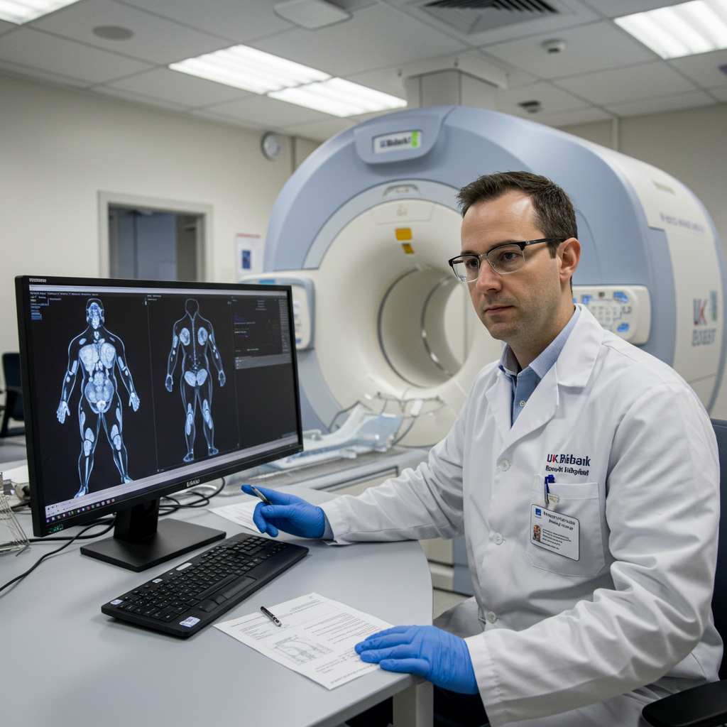
Scale and Scope of the Scans
The project scans multiple key organs in each volunteer. This includes the brain, heart, and major blood vessels. It also covers bones and joints. These detailed images provide a snapshot of the body’s internal state. They capture structural and functional details.
Each scanning session is thorough. It takes about five hours per volunteer. Participants undergo several different types of imaging. This typically involves multiple MRI scans, X-ray, and ultrasound imaging, including detailed scans of the neck’s blood vessels. This comprehensive approach gathers diverse visual data.
Context within the Broader UK Biobank
The true power of the imaging data comes from its integration. It is combined with the extensive information already in UK Biobank. This includes genetic data from DNA samples. It also incorporates lifestyle questionnaires collected over years. Physical measurements and health records are also linked.
This combination creates an unparalleled resource. Researchers can explore complex relationships. They can see how genetics, lifestyle, and environmental factors relate to internal body changes. This integrated view is key to understanding health and disease progression. Professor Sir Rory Collins, UK Biobank’s chief executive, highlighted this point. He stated that combining diverse data types provides a much better understanding of how bodies work. It also shows how diseases progress over time.
Why is This Study So Important for Research?
The detailed, large-scale imaging data offers unprecedented research opportunities. Studying 100,000 people provides statistical power. This allows scientists to identify subtle patterns. These patterns might link early physical changes to later health outcomes.
Unprecedented Detail for Research
The resolution and depth of the scans are highly detailed. They show minute structures within organs. Researchers can measure organ size, shape, and composition precisely. They can also assess tissue health and function. This level of detail was previously unavailable on such a massive scale.
Connecting Imaging to Other Data
Linking imaging data with other Biobank information is revolutionary. Scientists can test hypotheses about disease causes. Does a specific genetic variant correlate with a certain brain structure change? Does a particular diet affect fat accumulation in organs? The data allows exploration of these questions.
This resource supports data-driven discovery. Researchers can identify potential biomarkers. These are measurable indicators of disease or health. They can find early “warning flags” for various conditions. These might appear years before symptoms emerge.
Potential for Early Disease Detection and Prevention
A primary goal is finding ways to detect diseases earlier. Identifying signs before symptoms helps. It could allow for earlier interventions. This could involve targeted treatments or lifestyle changes. Professor Naomi Allen stated that researchers are using the data to identify disease early. They aim to target treatment at an earlier stage.
The data is also crucial for preventing common health problems. It helps researchers understand disease mechanisms better. This knowledge can inform public health strategies. It can also guide the development of new therapies. Conditions studied range from heart disease and cancer to dementia and Parkinson’s.
Behind the Scenes: The Scanning Process
The imaging project runs continuously. It operates 13 hours a day, seven days a week. Four dedicated sites across England host the scanning facilities.
Participant Experience
Volunteers like Steve, the 100,000th participant, play a vital role. Steve was motivated by his family’s health history, including his mother’s dementia diagnosis. He wanted to contribute to research. Participants undergo a multi-hour appointment. They move between different scanning machines.
While the process is detailed, it’s designed to be manageable. Volunteers contribute their time and data selflessly. Their participation is key to the project’s success.
Scanning Locations and Schedule
The four imaging centers are strategically located. They provide access for participants across England. Operating long hours ensures a steady flow of volunteers can be scanned. This maximizes the use of the expensive, specialized equipment.
Types of Imaging Used
A variety of imaging techniques capture different aspects of the body.
MRI (Magnetic Resonance Imaging): Provides detailed images of soft tissues and organs. Multiple types of MRI capture different properties.
X-ray: Used for visualizing bones and assessing bone density.
Ultrasound: Utilized for dynamic imaging, like assessing blood flow in vessels.
This multi-modal approach yields a comprehensive view of internal anatomy and function.
Managing the Data: Scale and Accessibility
The data generated is enormous. It currently exceeds 30 petabytes. That’s equivalent to 30,000 terabytes. Storing, managing, and making this volume of data useful is a significant technical challenge.
Massive Data Volume
Handling petabytes of detailed medical images requires robust infrastructure. High-performance computing is needed for processing. Advanced databases are essential for organizing and cataloging. This scale of data collection is unprecedented in human imaging studies.
Anonymization and Privacy
Participant privacy is a top priority. All data is anonymised before being shared with researchers. Identifying information like surnames or precise addresses is removed. Volunteers do not receive individual feedback on their scans. This is unless a serious, potentially life-threatening issue is incidentally spotted by radiographers. The project adheres to strict data protection protocols. Data security is constantly reviewed.
Open Access Model and Cost
Unlike some other large health databases, UK Biobank data is widely accessible. Researchers globally can apply for access. This includes scientists from universities, charities, governments, and the private sector. Access is granted at a low cost. Fees typically range from £3,000 to £9,000. This helps cover the running costs of the resource. This open access model fosters collaboration. It accelerates scientific discovery worldwide. Over 20,000 researchers in more than 60 countries currently use UK Biobank data.
Transforming Research: The Role of AI
Analyzing images from 100,000 people manually would be impossible. Professor Louise Thomas of the University of Westminster calculated it would take thousands of years. Artificial intelligence (AI) is essential for processing this massive dataset efficiently.
AI for Rapid Data Processing
AI algorithms are trained to analyze the images automatically. They can measure organ volumes, detect abnormalities, and extract complex features. Tasks that took manual experts minutes or hours can be done in seconds or minutes by AI. This allows researchers to process the entire dataset quickly. It unlocks the potential buried within the images.
One AI tool trained on Biobank heart images is now used globally. It analyzes heart scans in seconds. This task previously took around 15 minutes manually.
Examples of AI-Driven Findings
AI has already facilitated significant discoveries using Biobank imaging data.
AI combined with MRI and health data predicted the early onset of 38 common diseases.
Fat stored in muscle, detected by AI analysis of scans, is linked to age-related muscle loss and fall risk.
Analysis revealed potentially unhealthy levels of fat in the livers of up to a quarter of the UK population. This contributes significantly to liver disease.
Impact and Findings So Far
The UK Biobank, powered increasingly by its imaging component, is a research powerhouse.
Number of Publications
Since the project began in 2003, nearly 17,000 peer-reviewed papers have used Biobank data. Dozens more are published every week. The imaging data alone has contributed to over 1,300 studies in the decade since it began.
Specific Research Examples
Beyond the AI-driven findings, the imaging data has revealed critical links:
Combining MRI scans with other health data and an AI model can predict the early onset of 38 common diseases.
Even small amounts of daily alcohol consumption show a link to increased risk of memory loss and dementia based on brain imaging.
Detailed MRI scans might replace surgical procedures for diagnosing and monitoring a common form of liver disease.
Changes in heart structure, visible in scans, are associated with an increased risk of psychiatric disorders, including depression.
Techniques developed using Biobank MRI data are improving dementia diagnosis in NHS memory clinics.
Funding and the Future
The UK Biobank is a non-profit initiative. It relies on funding from major research bodies and charities.
Key Funders
Primary support comes from:
Medical Research Council (MRC)
Wellcome Trust charity
British Heart Foundation (BHF)
Department of Health
Scottish government
Additional funding for repeat scans has been provided by Calico (an Alphabet subsidiary) and the Chan Zuckerberg Initiative.
Plans for Repeat Scans
The project isn’t stopping at 100,000 initial scans. Plans are in place to rescan 60,000 of the participants. These follow-up scans will occur every few years. This creates a longitudinal dataset. It allows researchers to track changes in the body over time. This provides crucial insights into aging and disease development trajectories.
Frequently Asked Questions
What makes the UK Biobank imaging study so significant?
The UK Biobank imaging study is significant due to its sheer scale and depth. Scanning 100,000 participants with multiple advanced imaging techniques (MRI, X-ray, ultrasound) creates the world’s largest human imaging dataset. Crucially, this imaging data is linked to extensive genetic, lifestyle, and health record information from the same individuals. This unique combination allows researchers to study the complex interplay of factors affecting health and disease development in unprecedented detail.
How does the UK Biobank imaging data help researchers globally?
The anonymised imaging data from the UK Biobank is made available at low cost to researchers worldwide. Scientists from universities, charities, governments, and private sectors can apply for access. This open access model democratizes the use of this powerful resource. It allows diverse research teams to work on finding new ways to prevent, diagnose, and treat common diseases faster than if the data were restricted to a single institution or country.
What notable health discoveries have come from UK Biobank imaging?
UK Biobank imaging data has already led to important discoveries. Researchers have used it to show links between daily alcohol intake and dementia risk, and how changes in heart structure might predict psychiatric disorders. The data, often analyzed with AI, has helped predict early onset of diseases and identified potential biomarkers. It has also contributed to developing improved diagnostic techniques, like AI tools for faster heart scan analysis used globally or methods adopted by NHS memory clinics.
Conclusion
The UK Biobank’s imaging study reaching 100,000 participants is a landmark achievement. It transforms our ability to study human health on a massive scale. By combining detailed scans with rich biological and lifestyle data, scientists are unlocking secrets to disease prevention and early detection. This unparalleled resource, openly accessible to global researchers, powered by advancements in AI, is already yielding critical insights and paving the way for future breakthroughs in health and medicine. The commitment of volunteers and funding bodies has created a powerful engine for scientific discovery, promising a healthier future for generations to come.